
Home Microscopy Laboratory for Photomicrography
by Dr. Robert Berdan
February 3, 2018
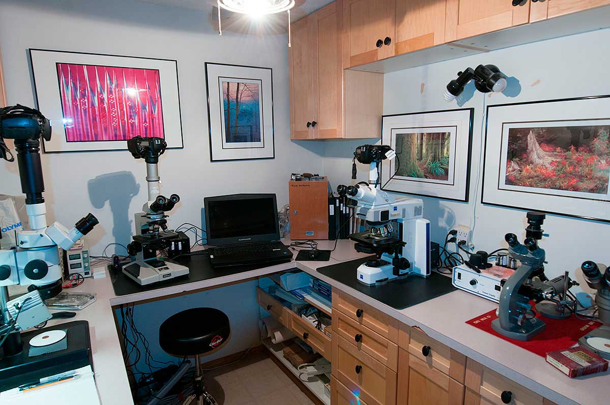
My darkroom converted into a microscopy laboratory for taking photomicrographs.
For the past 12 months I have been planning and building a microscopy lab in my home. It's about the geekiest thing a person can do I suppose and I am proud of it. Originally the small space I converted into a lab was a wine storage room and with the help of my father some years ago I refurbished it with counters and cabinets for use as a darkroom. My dream of having my own darkroom was short lived when in 2000 Epson produced the first archival ink-jet printers. With regret I sold my darkroom enlarger and accessories. From there on I processed my digital images on the computer using Photoshop and made prints with an Epson ink-jet printer. While I enjoyed working in a darkroom my skin had become sensitive to the chemicals and it took a long time to produce a few high quality prints. I was sad that 20 years of darkroom knowledge would no longer be of much value. Today most of my printing is done by photography outlets (e.g. London Drugs, Vistek) as they do a good job for less cost then printing myself. I still print some of my own exhibition prints, but if I don't use my Epson printers regularly the ink dries up and is unusable, and ink for Epson printers is expensive. The bottom line is my home darkroom has now been converted into a home microscopy laboratory (shown above) and I have just added a new Zeiss Axioscope Microscope with DIC optics - another dream come true for me. (DIC - Differential Interference Contrast also called Nomarksi after Georges Nomarski a Polish physicist who in 1952 developed this microscopy technique for Zeiss - Download PDF).

My Zeiss Axioscope microscope has two low power (2.5X and 5.0X) objectives which allows me to photograph entire arthropods. Above is Pediculus capitis - head louse viewed in bright field microscopy. 25X

Cross section of a leaf at low magnification. I produced this image by stitching several images together. 100X bright field microscopy.
When I was 10 years old my passion for biology and photography was ignited by a toy microscope - a Sears $29.99 model shown below. It wasn't long after receiving this toy microscope that I wished I had a way to photograph what I saw and share it with others. My first attempts started after I received a better microscope and purchased a Polaroid ED 10 - camera (see below).

My first microscope was from Sears and cost $30. It came with a carrying case, some slides, dissection instruments and offered 900X. I found this one recently on E-bay selling for $30 as a "Vintage" microscope - it now sits on my shelf as a reminder of where my passion for microscopy began.
When I think of what the early scientists had to use for microscopes in the 1600's and 1700's; for example Robert Hooke and Antonie van Leeuwenhoek this toy microscope doesn't seem so bad. However, I would not recommend it for anyone over 10 years of age, you would be better off buying a used research scope or one made in China or India for the young scientist. Nevertheless this toy microscope changed my life and lead me to a career in cell biology and neuroscience research.
I love working with microscopes and cameras. They bring my attention to living things that most folks will never see. The microscope and camera are two important tools that in my life have increased my appreciation for other life forms and my own.

Above is an advertisement for a Polaroid ED-10 camera for photomicrography that appeared in Scientific American. It was the first camera I purchased to take photomicrographs. The camera cost $70, had no lenses, just a shutter and an adaptor to fit on the microscope eyepiece. While it worked, the Polaroid print film was costly (for a teenager) and the prints were small. To share the pictures with others I had my prints copied into slides. Later on I received an Olympus OM-1 camera for Christmas and I began taking pictures using Kodak photomicrography film, Kodachrome, and Technical Pan black and white film that I developed myself. My father taught me how print in colour and I made 16 x 24 inch prints to hang on the wall. Most folks I showed the microscope images to didn't know or understand what they were looking at and showed little interest or appreciation for them, but I was not deterred. To my surprise I still sell some of the images of Diatoms I took over 40 years ago.

Above is an example of a student microscope and its basic parts. This microscope made in China costs about $300 new and about $100 used. It is a basic bright field microscope, but it can be easily fitted with polarizing filters, dark-field filters, Rheinberg Filters and would be good for most students to get started in microscopy. This scope is good for viewing prepared slides, histology slides and some microorganisms like yeast. Used microscopes like this are common on E-bay, Kijji and government surplus auctions. If you are lucky you may even find a used microscope with phase contrast for just a few hundred dollars.
When I graduated from Grade 8 I had saved about $150 and my parents contributed the rest in order to buy me an Olympus E microscope with a trinocular head for photography. I was fortunate that a close friend of our family, Herbert Thony, was a microscope salesman for Olympus and he sold me a microscope at cost and taught me how to setup phase contrast. Herb is now retired and over 90 years old. I visit Herbert when I go home to see my parents in Penetanguishene, Ontario. I dedicated my Master's Thesis to Herb as I am grateful for his help and also to my parents for helping me purchase the scope. Below is self portrait showing my first laboratory in our garage - I had this lab while I was in high school and through my undergraduate years at the University of Western.

My father built me a desk out of plywood, my Olympus E microscope is to my left and some of my photographs of mosquito larvae, potato starch grains, rotifers and copepods are hanging on the wall.

Above, my wife Donna (accountant and musician - not a scientist) used to to visit me in my University of Alberta Labratory since I spent most of my time there. I am lucky she was tolerant of my obsessive behaviour which seems to be a requirement to be a research scientist. She is sitting in front of an Olympus Inverted microscope IMT-2 which I modified to have a stereomicroscope above it, and it had an attached 35 mm camera (Olympus OM-4) and video port for time lapse videography. This inverted microscope had both phase contrast and DIC optics so I could film living neurons growing in culture. I used special micro-manipulators to impale the brain cells in order to record their electrical activity as they formed electrical synapses during growth and regeneration. The entire room was electrically shielded. While research was fun, there was constant pressure to be working in the lab and to publish more and more papers in order to get more grant funding. My problem is that I am interested in many other things, art, music, the outdoors, kayaking, computer technology, nature photography and spending time with my family. These are some things that many researchers have to sacrifice - some are willing to and others like myself were not. I don't believe we get a second chance at life and if you really want to do something you need to make it a priority.

Plant leaf stoma - 400X Polarized light microscopy - Nikon Optiphot microscope.

Cross section of pine wood showing the tracheids where water is transported up the stem of pine tree. DIC microscopy 200X, The pattern appears abstract but it provides important information to tree biologists.
About 18 months ago my passion for photomicrography returned. I have been photographing nature and wildlife around Calgary for over 25 years. There are still a few animals I have not captured on camera, however, I seemed to be spending more time looking for subjects that I already have pictures of. I started to look through the microscope and was reminded of just how many different organisms live in a small pool of water. I began exploring different optical techniques like Rheinberg lighting, Polarized light, Wave plates, Quartz wedges, Darkfield, Hoffman modulation, fluorescence and newer techniques and was drawn back to my first passion. I refurbished several used microscopes, and concluded that this might be my last chance to buy a research quality microscope with DIC optics to take pictures.
On the Internet I have been reading about other new advances in microscopy: Light sheet microscopy, confocal microscopy, Stimulation Emission Depletion microscopy etc. and I was amazed to read that the resolution of the light microscope is no longer limited to 0.2 microns, but that by using lasers (STED Stimulated emmission depletion microscopy) it is now possible to detect single molecules and the researchers that developed this technique received the Noble prize in 2014. The use of software to simulate different microscopy techniques is farily new and I am trying to get a hold of software from a researcher in Australia that can simulate Phase contrast and DIC using an ordinary bright field microscope and stacking the images (Download PDF on Optical Phase Microscopy). I have also been experimenting with focus stacking to enhance depth of field using digital photography and various software programs. To me the history of microscopy and photography are also fascinating topics (e.g. Download Robert Hooke's Micrographia 1664 -PDF). Hooke was the first to describe cells in Cork with his microscope). Also see some early photomicrographs of Diatoms by Dr. Redmayne from 1877 on this web site.

Diatoms are single celled algae that form glass (silica) shells (frustules) that can be highly ornamented. These single cells plants are believed to produce about 20% of the earth's Oxygen and some of them can move on their own - blurring the line between plants and animals. They can be found in most bodies of water and soil and Diatomaceous earth deposits left by millions of years of these organisms is mined and used in a wide variety of products like toothpaste, silver polish, beer filters, insect repellants and nano-technology. 100X DIC microscopy, Focus stack of 4 images from a prepared slide.

Diatoms DIC microscopy showing detail in the silica shells. Diatoms are also used to test the resolution of microscopes. 200X focus stack of 5 images.
When my son was born I decided to leave research. I was working 7 days a week 12-16 hour days and though I loved research I wanted a change. Success rates for grant funding by scientists at the time were less than 13%. At first I worked as manager of science education and exhibit development at the Calgary Science Center for 5 years as I needed a steady income for my family. I left in 1996 to start my own business "Science & Art Multimedia" where I combined my interests in Science, Technology, Art and Design. After 20 years my mortgage was paid off, and I have decided to focus on photography and microscopy. Fitness also is now a priority. My plan for the future is to show that Photomicrography can be considered an Art form as well as tool for discovery.

Cross section of a pine needle with DIC microscopy 100X
With a microscope you don't have to go further then your own backyard to find interesting creatures to photograph. Furthermore anyone can read about microorganisms online - research papers are readily available, and most scientists reply to emails and seem to be glad to share their research findings. Lab supplies, and microscope parts can be purchased for reasonable prices on Kijjii, and E-bay and Amazon sells almost everything. What an exciting time!

Cross section through a pine needle with DIC microscopy, 100X The background colour can be changed using the DIC prisms and by using a full wave plate. The wave plate also works with a polarizing microscope.
Anyone can start taking pictures through a microscope with a simple camera adaptor that sells from $20-$100 and there are also a wide variety of phone adaptors to connect to your microscope. If you do plan to use a microscope to take pictures it helps to know some science and microscope fundamentals. If you plan to buy a microscope - I recommend you seek advice of an experienced microscopist (you can email me). Many of the older microscopes (10-40 years) if cared for are excellent for taking pictures. Also , you don't have to take pictures with your microscope, just start exploring the world around you. Children can get started with a simple magnifying lens to stimulate their curiosity about plants and animals. If you are new to microscopy I recommend you buy a used microscope under $500 at first and then if the microscope ignites your passion you can always sell your scope and buy a better model.

Cross section of a corn stem (Monocot) - stitch of several images 20X DIC microscopy

Part of a cross section of a plant (monocot - Corn) shown above viewed with DIC microscopy. 200X

Cross section of plant tissue showing spherical objects - starch grains 200X DIC microscopy.
When I decided to purchase a research quality microscope with DIC ( Differential Interference Contrast also called Nomarski), I already had several microscopes, but DIC adds colour and 3 dimensional appearance to living specimens. Unfortunately even a used microscope with DIC can cost about $10,000. I pondered how I could afford a DIC microscope and still stay married? I figured out that if I sold some of my camera equipment and other belongings I might be able to raise enough money. So I put my telescope, alto saxophone, two kayaks, several lenses and other accessories for sale on Kijji. By November 2017 I had raised about 50% of the money I needed and decided to purchase a Zeiss Axioscope microscope with DIC and phase contrast optics. The Zeiss sales person was kind enough to find me a demo model and offered me a discount - thanks Lita! Now that I have a microscope with DIC - I haven't been this excited in years.

Citric acid crystals DIC microscopy 200X

Mineral section viewed with DIC microscopy 200X
When my new Zeiss microscope arrived in January it was a dream come true. I am planning to share my new photomicrographs in the coming months- this is the first article. I will be photographing freshwater invertebrates like water bears (Tardigrades), ciliates, rotifers, copepods, ostracods, coelentrates (hydra), crystals, pollen, plant sections and more. My hope is that my photographs might inspire some readers to try microscopy and photomicrography and gain a better appreciation of the microorganisms around us. If you are an arm chair scientist you can just check out the articles and pictures on my site and others. In the UK and Europe microscopy has a long history and as a hobby is far more progressive than in North America where microscopy is used mainly by research students and scientists - it doesn't have to be. Microscopy can be a gateway to new worlds without leaving your backyard - you will be transported to another dimension of the very small. For me microscopy is more then just capturing pretty pictures - I am also learning more about many of the Freshwater invertebrates and their importance in our ecosystem which we are all a part of.

Above are human cheek epithelial cells (mine) shown with different microscope techniques. A) Positive phase contrast 400X B) Negative Phase contrast 400X c) DIC - Nomarski 400X D) Hoffman modulation 1000X. Living cells are transparent so in order to view live cells you need to either stain the cells or use a microscope with special optical contrast techniques. If you only have a bright field microscope you can also use stain, e.g. methylene blue, or food colour etc to impart colour to the cells.

Plant cells (onion) stained, nuclei are red and cell walls appear blue - using DIC microscopy 400X

Radiolaria - these one celled organisms live in the ocean and like Diatoms they form intricate silica shells. 400X focus stack 4 images DIC microscopy.

Above in the foreground is my new Zeiss Axioscope microscope with my Nikon D800 camera attached on top connected by a USB cord to my laptop. I am using free software to capture the images called Digicam control. My Zeiss microscope includes several objectives: 2.5, 5, 10, 20, 40 and 63 X. These objectives can be used with polarized light, phase contrast and DIC (differential interference contrast), Rheinberg filters etc. I am currently in the process of adding Phase contrast objectives - 10, 20, 40X to the Axioscope. In the background is a Nikon Optiphot microscope which I purchased from Kijji and I have been refurbishing for about year now. It's a 20 year old research microscope that uses polarized light and flourescence illumination but it doesn't have the DIC optics of my Axioscope.

50 years later I am still a science geek. On the right is my first Olympus E research microscope I received in Grade 8 , and in the background are some pictures and graphics from my cell and neurobiology research. This photo was taken by my friend Dr. Sharif Galal. In the near future I hope to share more information and pictures of freshwater invertebrates and various microscope technologies. Microscopy is something anyone can enjoy and it might even encourage some younger folks to enter careers in science.
References and Links:
Zeiss microscopy Canada - online store
Microscopy from the very beginning - Ziess PDF
Quality Microscopes Ltd - specializes in stereo microscopes for Petrology and is located in my neighbourhood
Nikon Microscopy - professional research quality scopes
Olympus Microscopy - research quality scopes
Olympus microscopy resource center
Leica Microscopes - research and educational microscopes
McCrone Microscopy Group - Chicago, learning center, also has wide variety of articles on microscopy
The Quekett Microscopical Club England - lots of great articles on how to get started in photomicrography
Microscopy-UK Miscape Magazine - many articles on all aspects of microscopy for all levels
Brunnel Microscopes Ltd UK & International specialists - wide variety of hard to get accessories and scopes
The Royal Microscopical Society since 1839 - UK
Microscopy Today Online - Free online magazine about new trends in microscopy
Live Cell Imaging Microscope Resource Lab - University of Calgary - guest speakers, learning programs etc
Boreal Science - science teaching resources, you can purchase live protozoa, supplies etc .
The Microbehunter - Setting up a Home Laboratory for Microscopy
Setting up a Home Chemistry Laboratory
AM microscope in California - microscope adapters, microscopes, accessories - they offer good service and prices
Valley Microscope - Ottawa Ontario, they sell student microscopes and prepared microscope slides
Vermont Optechs - US based offer very reasonable priced used research quality microscopes
Radical microscopes from India - they offer microscopes and accessories for reasonable prices.
How to get Chemicals for a Home Lab - Youtube Video
Lab Microscopes on E-bay - you can get some good deals, but do your home work and research before you buy!
Setting up a home science lab
Doing Biotech in a Bedroom
Safety tips for setting up a home chemistry lab
MSDS (Material Safety Data Sheets) if you are using chemicals know the dangers.
Download "Ernst Haekel's Artforms in Nature" - beautiful hand drawn Radiolarians - Free PDF
Acknowledgements: I would like to thank Lita MacDonald the regional Zeiss sales representative in Alberta, Sarah Croteau Sales coordinator at Zeiss in Toronto, and John Chan (technical engineer) from Zeiss microscopy. Doug Hayden at Quality Microscope sells Zeiss stereo microscopes and provided me with some components and advice.
Authors Biography & Contact Information

Robert Berdan is a professional nature photographer living in Calgary, AB specializing in nature, wildlife and science photography. Robert offers photo guiding and private instruction in all aspects of nature photography and Adobe Photoshop training.
Email at: rberdan@scienceandart.org
Web site: www.canadiannaturephotographer.com
Phone: MST 9am -7 pm (403) 247-2457.
Click on the buttons below and share this site with your friends