
Photography beyond the visible
Come along with me on my travel through the microcosm...
by Gerd Günther
February 5, 2019

My name is Gerd Günther, I was born in Düsseldorf, Germany in 1958; after my schooling days the education in agricultural sciences at the University of Göttingen followed. Since 1986, I have been working as self-employed farmer in Düsseldorf.
Motivated by my teachers in school, I got into photomicrography 45 years ago. At that time we experimented with analogue film material in black-and-white with poor results. With the upcoming of the digital era photomicrography intensified continuously since 1998. Photomicrography is my passion. In order to dive into the microcosm, a tool is needed: a microscope.
For several years now I have been working with a Leica DMLB research grade microscope. Over the years I upgraded this machine with Epi-fluorescence contrast and differential interference contrast as well. The adaptation of digital cameras was realized with homegrown parts. Additionally an electronic flash is adapted via double-collector system to ensure sharp details of fast moving objects.
Exploring the microscopic world is hugely attractive, moreover travelling distances in a microscopic expedition are quite short. In animate as well as inanimate nature we can discover an overwhelming wealth of forms and structures, looking through the eyepiece of a light microscope helps to expand our horizon of experience, far beyond pure observation. As a "micronaut" you just have to step out your front door to enter the microscopic world. I think, everyone is overwhelmed by the broad bandwidth of creatures living in aquatic habitats. So let's begin with our travel through the microcosm right there:

Rotaria sp. rotifer. Rotifers are small animals, living in nearly every puddle and lake including the world's seven seas.
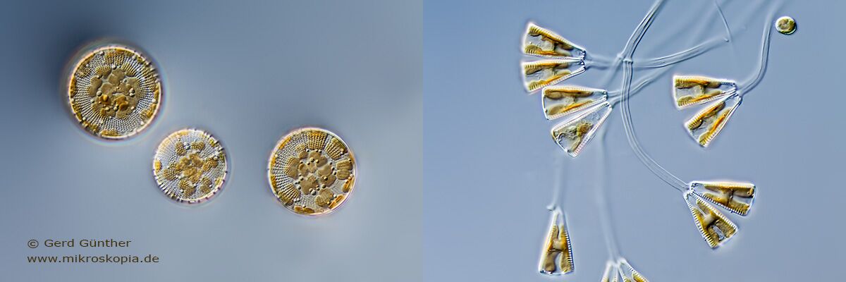
Staphanodiscus sp. diatom (left) and Gomphonema sp. diatom (right) belong to a group of single celled plants, living in a shell composed of glass, Silicon dioxide. Worldwide there are about 285 genera and 12,000 species recognized. Diatoms are very abundant primary producers, responsible for about 20% of the global carbon fixation. They produce as much oxygen as the land plants.
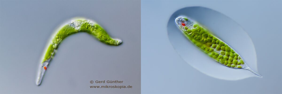
Euglena mutabils (left) and Lepocinclis tripteris (right) belong to the Euglenoids, a group of protists, they are photosynthetic species with chloroplasts and have a characteristic red eyespot.

Chaetophora (left) and Xanthidium antilopaeum (right) are aquatic green algae. Xanthidium produces masses of mucilage, surrounding each cell, shown here with the embedding in Indian ink.
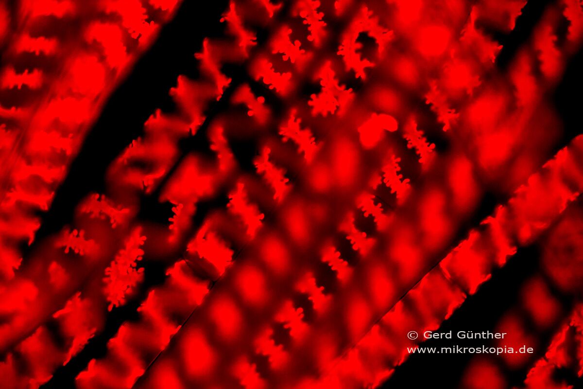
Epi-Fluorescence with blue excitation gives a wonderful deep red autofluorescence of the chloroplasts of this desmid, Spirogyra sp. This contrast method gives an interesting, more detailed 3D impression of this filamentous alga.

The dinoflagellate Pyrocystis noctiluca lives in warm seas and has the ability to show intense bioluminescence, wonderfully captured here : https://vimeo.com/29871792
Another group of organisms we frequently find in aquatic samples are ciliates, single-celled animals. There also is a wide bandwidth of species in freshwater and marine habitats as well. Ciliates are a bit more complicated in photomicrography due to their permanent, sometimes very fast movement. The use of an electronic flash is recommended here.
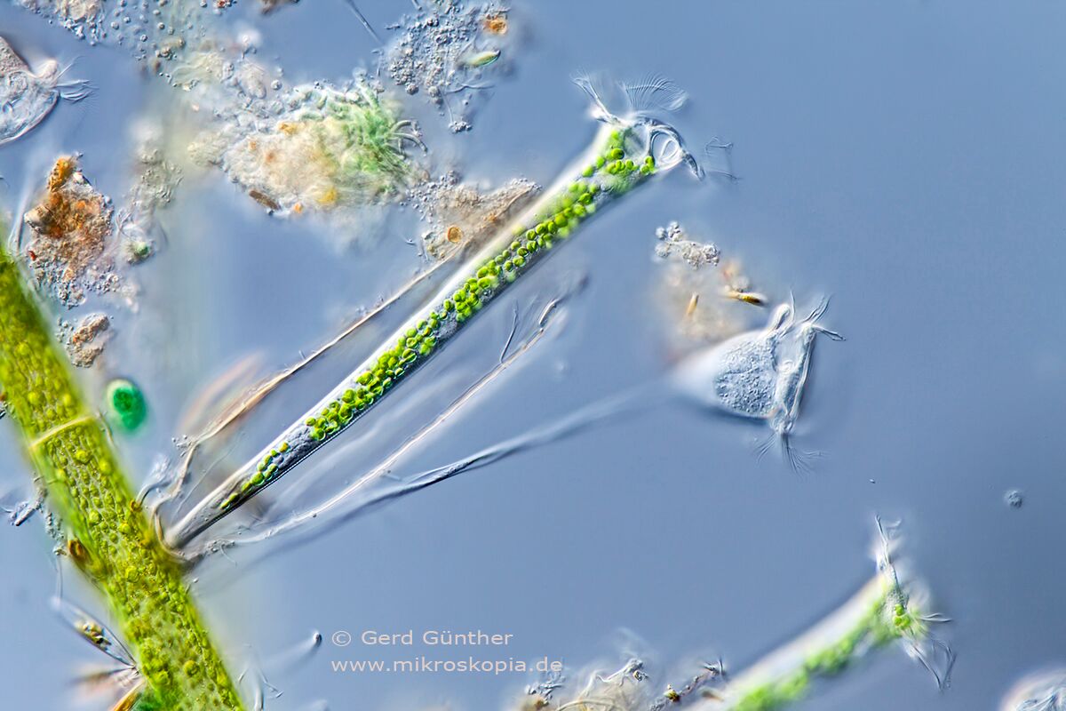
This is an image with peritrich ciliates, Thuricola folliculata with a lorica and green symbiotic algae, genus Chlorella together with bell-shaped ciliates, genus Vorticella on their flexible stalk.

Paramecium bursaria, a common hymenostome ciliate inhabits several green algae genus Chlorella and shows its sculptured pellicle when squeezed under the coverglass.

Loxophyllum meleagris is a huge, about 300 µm long predatory pleurostomatid ciliate with a moniliform macronucleus and lots of toxicyst warts.

This is a very special astomate ciliate, Metaradio sp., living commensally in earthworm guts. Often these ciliates occur abundantly, with nearly every worm infected. We wrote a small article in acta protozoologica which can be found here : http://www.ejournals.eu/Acta-Protozoologica/Volume-55-Issue-1/art/6453/
Exploring the world of plants is another wide field where you can find interesting details. The thin sectioning of plants is relatively easy, a razorblade is a sufficient tool to enter the world of plant anatomy.

Thin section through a leaf of Thymus vulgaris, common Thyme, showing the thick xeromorphic epidermis cells and one peltate trichome. Peltate trichomes produce most of the essential oil, with terpenes comprising the main component.
Dried onion skin in polarized lighting. Often simple samples out of the kitchen show an unexpected beauty when seen with a microscope.

All plant hairs and trichomes are worthy subjects, these stamen hairs out of an unopened Tradescantia virginiana flower are wonderful objects to show cell division in vivo. The cell at the tip of the left stamen hair divides into two new daughter cells, showing Anaphase stadium.

Unripe, green seed grain of Shepherd`s-purse, Capsella bursa-pastoris, taken from a wildflower. The small seeds are located in the purse-shaped seed capsule, containing up to 40 seeds.
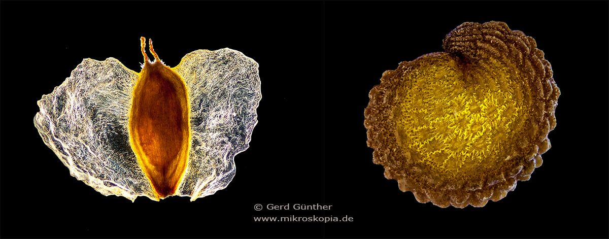
Seed grains of birch (left) and Common chickweed, Stellaria media (right)
When we accept a bit more of preparation input, we can explore the inner structure of many technical subjects under the microscope. The preparation includes embedding of the samples in light curing resin and then grounding and diamond polishing.

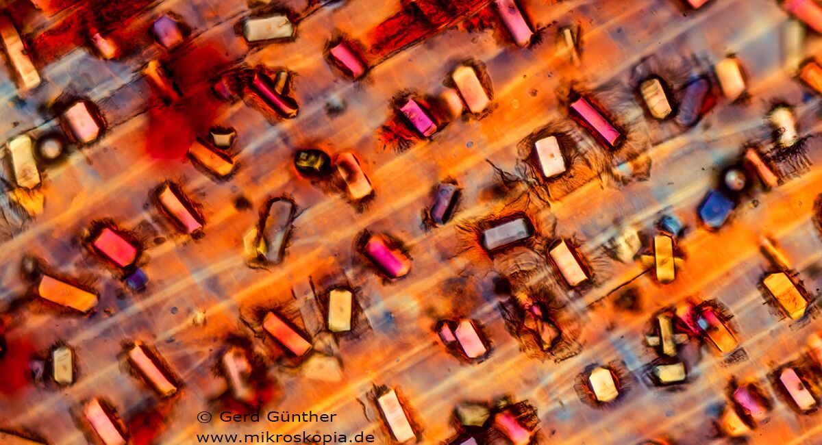
Section through a multilayered printed circuit board (PCB) from a computer. The image shows the basic epoxy material of the PCB with glass reinforcement filaments in linen weave (blue) with two copper layers of the multilayer.

Section through a computer printed circuit board (PCB) with surface mounted ceramic capacitors. The image shows the basic epoxy material of the PCB with glass reinforcement filaments in linen weave in blue color with two copper layers of the multilayer. Two ceramic capacitors in cross section are visible on the upper side of the PCB, showing the interior composition of the capacitors with several thin metal plates separated by ceramic material.
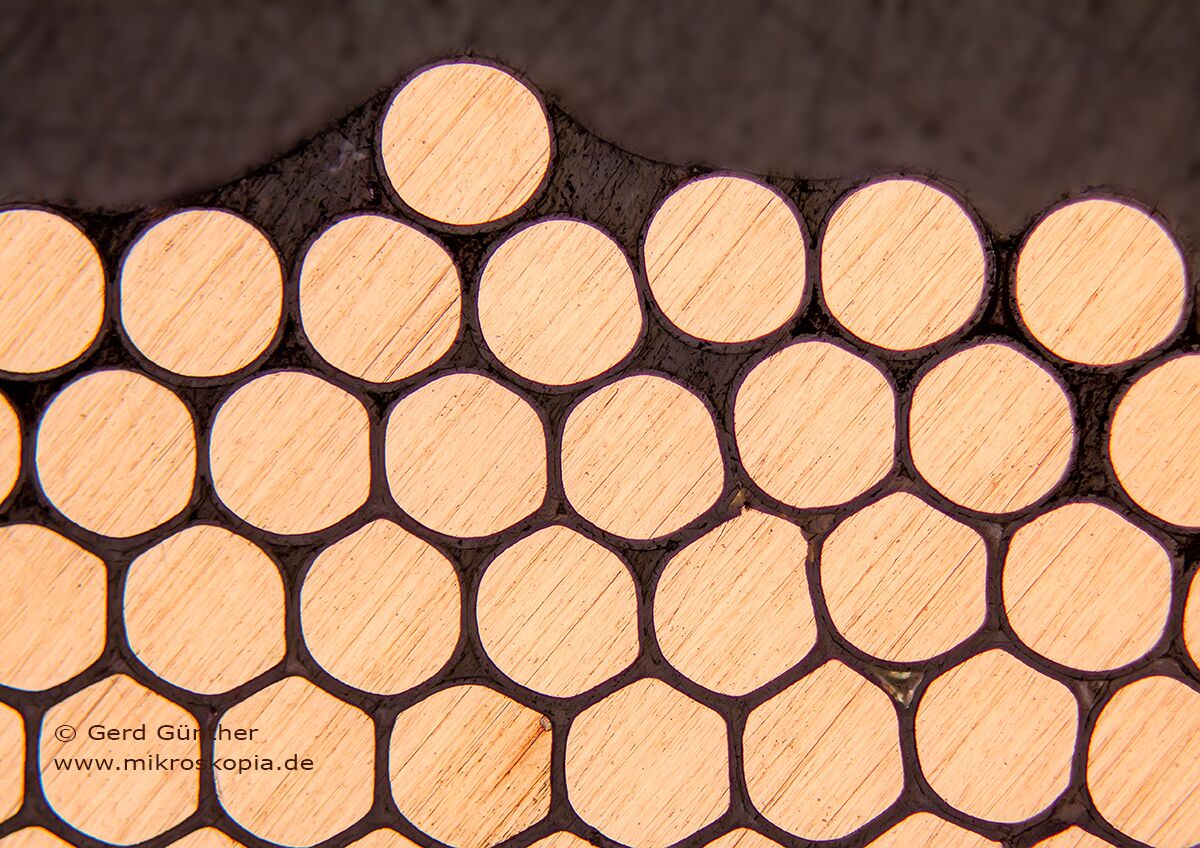
Longitudinal section through an inductivity, a solenoid used for printed circuit board mounting. In this orientation the copper wires are cross sectioned.
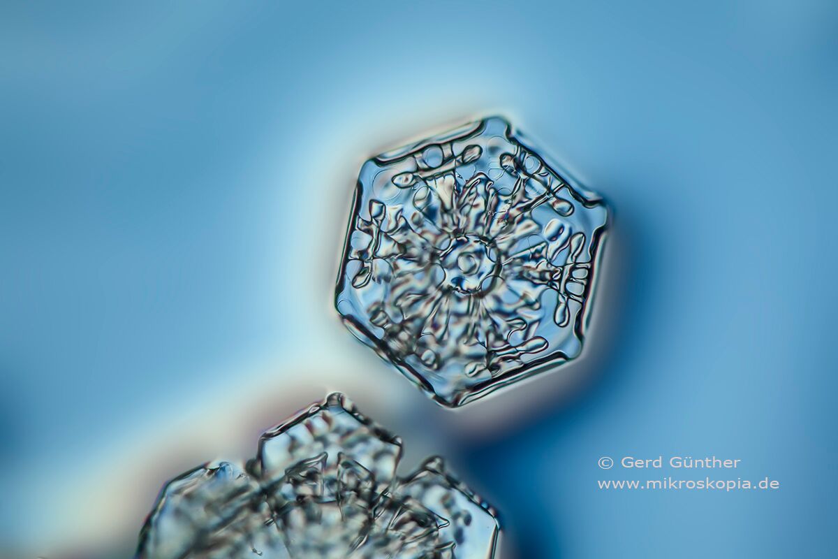
Snowflake, caught in a thin layer of nail varnish
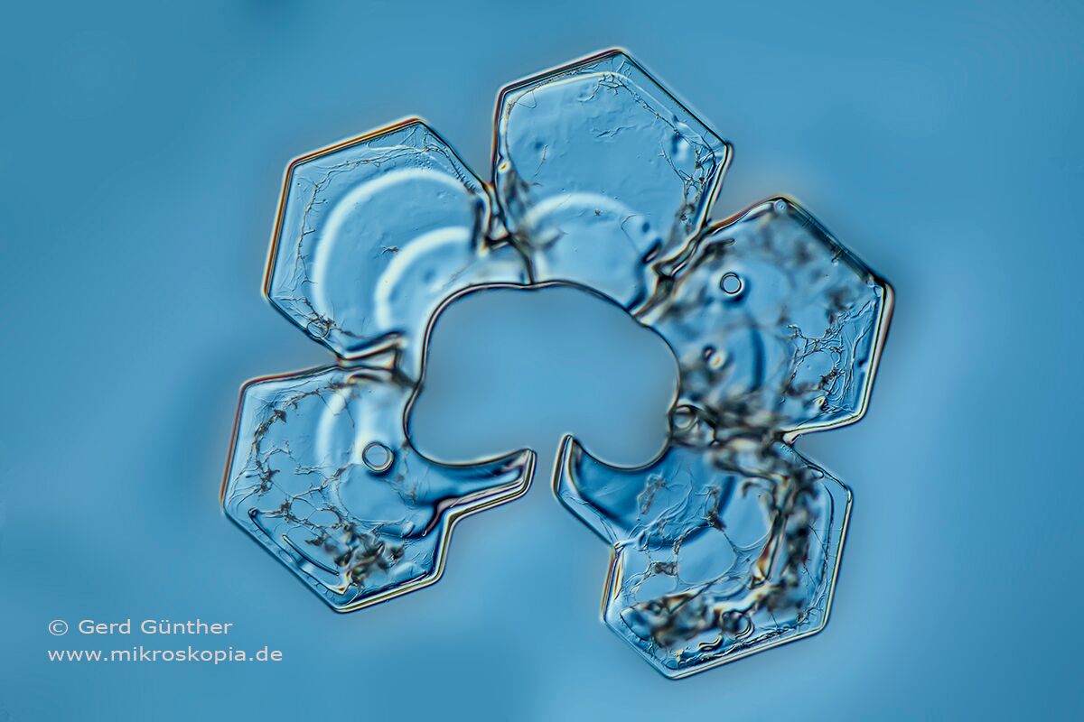
The snowflakes were conserved in a thin layer of nail varnish and captured with differential interference contrast.
Authors Biography & Contact Information

I work as a freelance photographer with emphasis on phycology, protozoology and plant anatomy.
Please contact me via my website: http://www.mikroskopia.de/contact-en.html
Website: www.mikroskopia.de
Instagram: https://www.instagram.com/mikrohiker/