
The Eye of the Beholder - What every photographer should know about their aging eyes
by Dr. Robert Berdan
Updated April 24, 2019
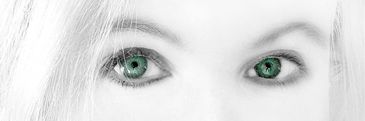
Beautiful Green eyes of my wife
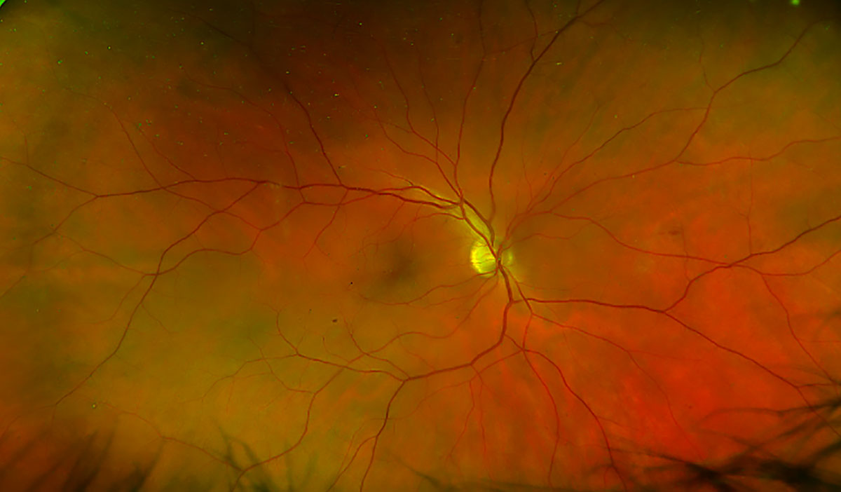
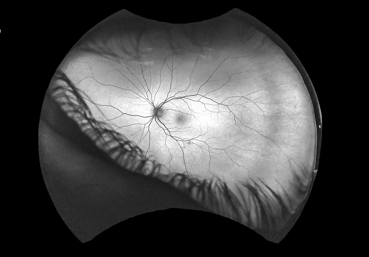
Retina photograph of my right eye showing blood vessels
Our eyesight is important in almost everything we do in our lives and to a photographer or a visual artist good vision is essential. As we age our eye sight gets poorer. Between the age of 20 and 60 the amount of light reaching our retina can decrease by two-thirds. In this article I share some insights in our eyes and how they change during aging. I also include some retina photographs taken during a recent eye examinations because I found the photos fascinating.
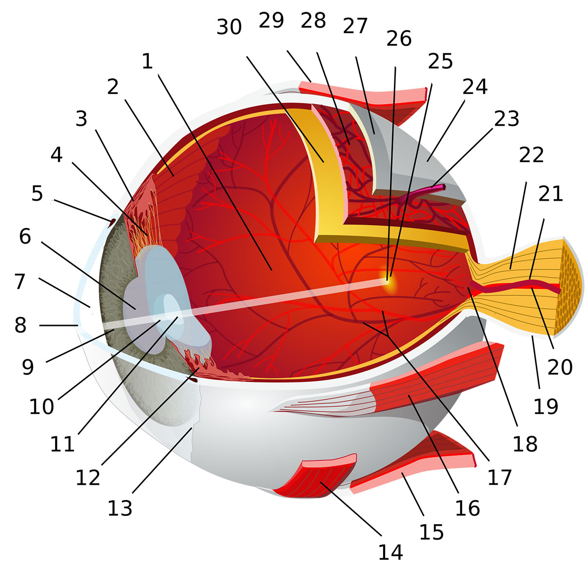
Diagram of the human eye 1:posterior segment of eyeball 2:ora serrata 3:ciliary muscle 4:ciliary zonules 5:canal of Schlemm 6:pupil 7:anterior chamber 8:cornea 9:iris10:lens cortex 11:lens nucleus 12:ciliary process 13:conjunctiva 14:inferior oblique muscle 15:inferior rectus muscle 16:medial rectus muscle17:retinal arteries and veins 18:optic disc 19:dura mater 20:central retinal artery 21:central retinal vein 22:optic nerve 23:vorticose vein 24:bulbar sheath 25:macula 26:fovea 27:sclera 28:choroid 29:superior rectus muscle 30:retina (diagram source Wikipedia).

Above cross section through an animal eye showing the retina and lens. About 10X

Section through the eye of a monkey showing the retina with inside of the eye at the top - DIC microscopy. The rods and cone cells are at the bottom. The entire retina is about 0.25 mm thick.
I receive regular eye checkups and during my last examination I was injected with sodium-fluorescein a dye I used for microinjection into cells when I was a graduate student studying cell-cell communication. The purpose of the retinal examination was to look for blood vessels in my retina that leak indicating damage to the blood vessels. Thankfully so far my eyes seem to be OK. I asked if I could get a copy of some of the pictures of my retina in order to share them as I thought it was interesting to see what we see with. All photographers should be aware that their vision will change as they age, and should protect the eyes primarily by having regular eye-checkups, exercise and eating fruits and vegetables and if doing things that might damage the eyes like cutting the grass, working with certain machinery - one should wear protective glasses.

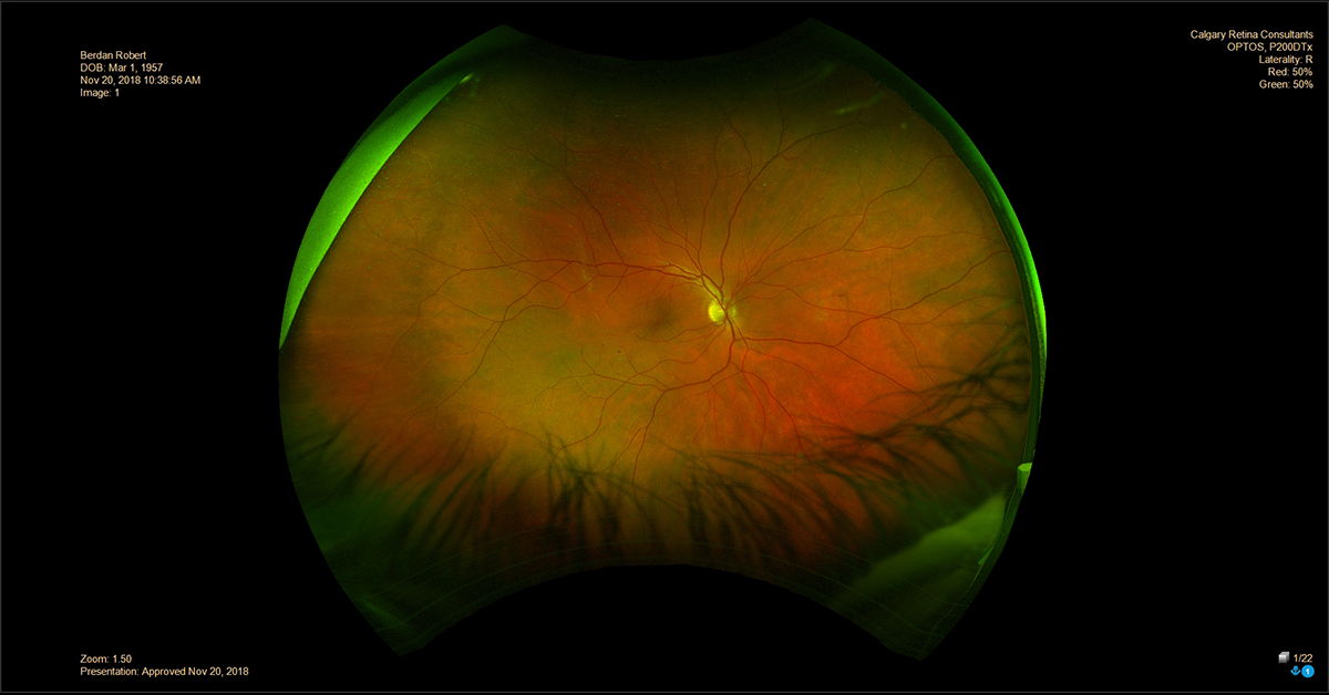
The retina of my left eye after being injected with a fluorescent dye - sodium fluorescein.

The retina of my right eye photo was taken in 2006 showing the blood vessels. My eyes were fine but I needed to start wearing glasses. The blood vessels converge on the optic nerve.

The retina of my left eye photo taken in 2006
As we age our eyesight generally declines, we need glasses to read and the retina of a 60 year old typically receives on the third the amount of light of a 20 year old. The cornea of the eye flattens limiting the ability to focus and may also become flecked with fatty deposits. The smaller opening of the pupil in older individuals, a condition called miosis, also limits the amount of light entering the eye. Just as a smaller opening in a camera lens reduces the amount of light, it also enhances depth of field and the one benefit is that some older folks may report an improvement in their eyesight in bright light. However our vision in dim light is significantly reduced.
Fundus Photography
Fundus photography documents the retina, the tissue at the back of the eye that translates optical images into electrical signals the brain interprets and understands. A fundus camera or retinal camera is a specialized low power microscope with an attached camera that can photograph the back of the eye or retina, including retinal vasculature, optic disc, macula and posterior pole (i.e. the fundus is the part of the eye opposite the pupil hence the name Fundus Photography).
Fluorescein Angiography is a procedure used to record blood flow in the retina. Fluorescein is injected into a vein in the hand and it travels throughout the body. You are then positioned in front of the fundus camera by resting you chin and pressing your forehead against a bar while the opthalmic technician photographs your eye. Photographs can reveal if any blood vessels are abnormal, leaking and damaged. Before having this procedure your eyes will be dilated with a chemical agent and you should avoid caffeinated beverages before this. The procedure is safe and does not cause injury to your eyes nor does it exacerbate any retinal degeneration (M.A. Mainster and P.L. Turner 2009), but your will urine will be fluorescent for the rest of the day and some individuals who might be allergic to Flourescein may exhibit some itchiness.
Smart Phone Fundus Photography
It is possible for doctors in emergency rooms or living in first-world countries to use a smart phone to photograph and diagnose optic nerve and macula pathology. The eye needs to be dilated with (2.5% Phenylephrine and 1% Tropicamide for 20 minutes) by a physician, and requires an accessory opthimoscopy lens, and smartphone used in video mode - to see how this is done see this video.
Flourescein angiograph of the back of my eye - you can see my eyelash on the bottom.

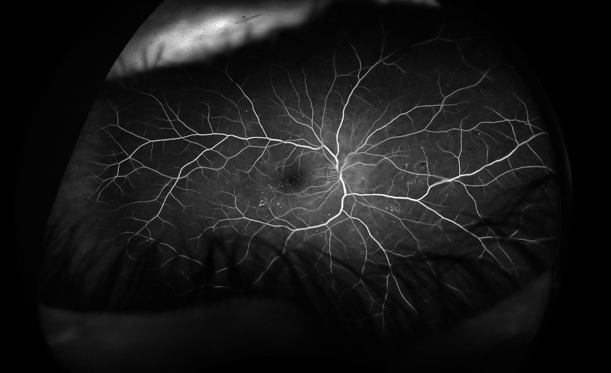
Black and white image of my right eye - showing blood vessels.
Black and white image of my left eye - showing blood vessels.

Image of my left eye showing Fluorescein in the branching blood vessels at the back of my retina.
Positive image of my right eye showing fluorescein in the blood vessels at the back of my retina.
Blue light is used to excite the sodium flourescein which glows a bright green colour or appears white in black and white photos.
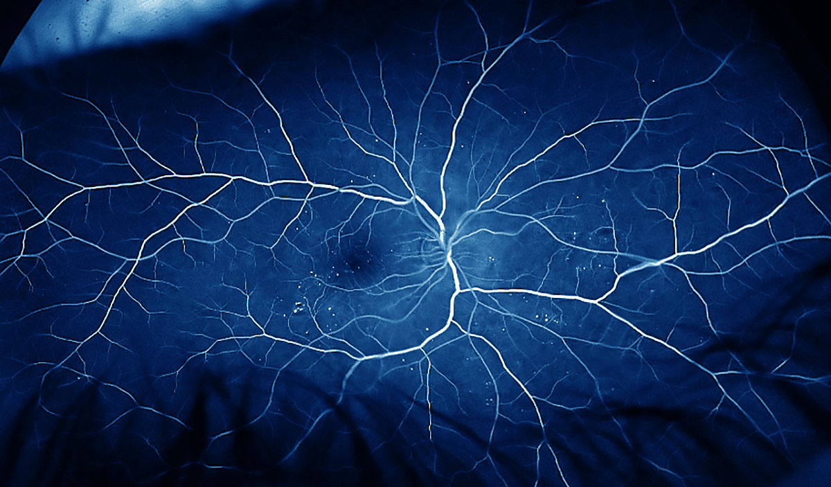
Above is a photograph of my right retina showing the blood vessels branching like a tree - image was toned blue in Adobe Photoshop.
Our cameras
Most digital cameras today have autofocus features which helps us acquire sharp focus as we get older, and the purchase of lenses with wide openings e.g. F2.8 make it easier to focus indoors or any place that has low light which is why I prefer lenses with an F2.8 opening most of the time. Photography of the night sky and aurora borealis are examples of low light photography.
When looking for the aurora you can increase your chances of seeing it early in the evening by using your peripheral vision which uses the rods located in your retina. We have many more rods in our retina and they are sensitive to low light. Cones are used for colour and concentrated in fovea and macula near the center of the eye. There are about 120 million rods throughout the retina whereas we have about 10 million cones. It is estimated that the human eye is equivalent to a 576 megapixel camera, has a dynamic range of about 20 or more stops and a focal length of about 22 mm (clarkvision.com), I have also read our eye has a focal length of 43 mm elsewhere. Our eyes process images at the equivalent of about have about 1\30 second shutter speed which is why movies or video recorded at 1\24 to 1\30 of a second appear smooth to us.
Healthy Eyes
Pure Optical says our risk of developing eye disease increases with age and certain conditions like diabetes or high blood pressure can make our eyes more susceptible to disease. It's also important to protect our eyes from ultra violet light by wearing protective glasses. Our general health and diet are important to having healthy eyes. Vitamin A deficiency is one of the most common causes of blindness in the world and not having enough can result in night blindness. Fortunately Vitamin A is found in high amounts in fruits and vegetables like spinach and carrots. Other elements that are found to be important are Lutein and Zeaxzanthin which are carotenoid antioxidants and may reduce the risk of macular degeneration. These elements are found in spinach, parsley, and egg yolks. Other important nutrients include Omega-3 fatty acids, Vitamin C, and Zinc (for more information see A. Arnarson 2016).
As we age our eyes will change, the brightness will decrease, our colour perception may change (maybe that is why I like to process my images with a more saturation then some folks care for), and some of us will need to wear glasses or contacts. Research studies have shown that most animals kept in confined environments became near sighted. When I was younger I had 18\18 vision and when I was 21 it became 20/22 after measuring about 100,000 particles using a loupe with my right eye. Apparently the strain of measuring the particles permanently changed my vision in one eye. What 20\20 vision means is that when you stand 20 feet away from an eye chart, a person can clearly identify a row of 9 mm letters from 20 feet. The best vision ever recorded was an Aboriginie man with 20/5 vision. For more information on 20/20 vision see commonwealtheyes.com.
As I get older I have noticed it's more difficult to read small text, and driving at night its more difficult to see and the glare of oncoming car lights is more annoying. There is not much we can do about aging other then exercise, eat healthy food and have regular checkups, but as photographers we should be especially aware of how our vision may change as we age.
References
The Aging Eye - National Center for Biotechnology information - download PDF
Dr. Anna Ells provided the recent photos of my retina - located at Southern Alberta Eye Center.
Dr. Bishop at Northland Mall provided two retinal photos of my eyes taken in 2006.
Fluorescein Angiography - Univ. of British Columbia
Fundus Photography Overview - shows a normal eye, Glaucoma and Diabetic retinopathy.
Notes on the Resolution and other Details of the Human Eye - ClarkVision.com
What's digital retinal photography? YouTube Video.
Macular Degeneration and the Aging Retina - YouTube Video.
Smartphone Fundus Photography - Video & PDF
8 Nutrients that will optimize your eye health
Authors Biography & Contact Information

Robert Berdan is a professional nature photographer living in Calgary, AB specializing in nature, wildlife and science photography. Robert retired from Cell\Neurobiology research to take up photography full time years ago. Robert offers photo guiding and private instruction in all aspects of nature photography and Adobe Photoshop training - including photomicrography, macrophotography.
Email at: rberdan@scienceandart.org
Web site: www.canadiannaturephotographer.com
Phone: MST 9 am -7 pm (403) 247-2457.