
Photomicrography of Rotifers II
Brachionus manjavacas and Philodina roseola
by Dr. Robert Berdan
March 12, 2020

Above Brachionus manjavacas stained with Acridine orange and viewed in a fluorescence microscope 400X.
Most folks are not aware of rotifers, yet probably walk by them daily. These tiny creatures live in a film of water within moss embedded on the sidewalk, lichen growing on a tree, in puddles of rain water, or even a bird bath. Rotifers also live in eves troughs, ponds, rivers, and ocean estuaries. You can't see these tiny creatures unless you have a microscope as most of these animals are only 0.10 mm to about 1.6 mm in length. Rotifers form an important link in the food chain providing nutrition for small fish larva, crustaceans and other small aquatic organisms. They feed primarly on bacteria, yeast and algae. Rotifers are also used in research to study aging, nutrition, and toxicology and there are no known harmful effects of rotifers on humans.

B. manjavacas rotifer viewed by differential interference microscopy (DIC) also referred to as Normarski contrast after the inventor Georges Nomarski. This technique provides a 3D relief of the specimens or cells and the background colours can be modified from grey to a variety of interference colours using a full wave retardation plate. Note the red eye and the yellow structures which are jaws (trophi) made of chitin. 400X.
What fascinates me about rotifers is that they come in a wide variety of shapes and have an alien-like appearance. Some rotifers can move like an inch worm, or use small hair-like structures called cilia around their anterior ends that appear to rotate like wheels - hence they were originally called "wheel-bearer" (from Latin rota “wheel” and -fer “bearing”). I have been photographing these minute Animalcules ("little animal", from Latin animal + the diminutive suffix -culum) for decades. Rotifers were first described by Rev. John Harris in 1696 in the Philosophical Transactions of the Royal Society. Other rotifers were described by Antonie van Leeuwenhoek in 1703 and also by Baker (1764). The microscope was invented circa 1590. Some historians credit Zacharias Janssen others credit Hans Lipperhey, though we may never know for sure who invented the microscope (Cox 2013). What's important is that the microscope opened up a previously invisible world and had a profound impact on medicine, biology and scientific research. Photomicrography began in the early 1800's (N. Overney and G. Overney 2011).

The Rotifera strictly speaking are confined to the Bdelloidea and the Monogononta. Rotifera, Acanthocephala and Seisonida make up a clade called Syndermata (Wikipedia). A clade is a group of organisms believed to have evolved from a common ancestor. The Monogononta are characterized by the fact that both male and female rotifers have only one genital gland, whereas the Bdelloidea have two glands and there are no males. Bdelloids reproduce without sex. The phylum Rotifera currently includes 2149 species (F. Leasi and W.H. De Smet 2020) with Monogononta having over 1700 species, while the Bdelloidea about 455 species. The Seisonida contain 4 epizoic marine species.

Wheel animacules drawings from Baker (1794) page 288 - showing a variety of Rotifers.
Before reading this article, you might first want to look at my previous article "Photographing Rotifers" on this web site. In this article I concentrate on two rotifers Brachionus manjavacas and Philodina roseola and present photomicrographs of them using a variety of microscopy techniques. At the end of the article I also include a few pictures of rotifers I found locally in Calgary, Alberta, Canada.

Brachionus manjavacas viewed by bright field microscopy 400X. A bright field microscope is the most common type of microscope and with it one can see pond organisms and bacteria. Other microscope techniques like phase contrast and Differential Interference contrast (DIC) show more cellular detail in living cells which normally appear translucent. These other microscope tools are also more expensive and found mainly on research light microscopes. The Brachionous and Philodina species shown here were kindly provided by Dr. Terry Snell a leading rotifer biologist in the US.

Brachionus manjavacas stained with Lugol's iodine (a solution of potassium iodide with iodine in water) which interacts with polysaccharides, starches and glycogen in animals cells viewed by bright field microscopy. 400X

B. manjavacas stained with Toluidine blue and viewed by Bright field microscopy. 400X

Female Brachionus manjavacas by DIC microscopy. Males are much smaller in size and rarer. Males have only one function to fertilize the eggs, most males can't even feed. The vitellarium where eggs are held in the female above is not visible in this view and neither is the foot and toes but see picture above and below this one. 400X

Brachionus manjavacas DIC microscopy 400X

Trophi or jaws are used to grind, pierce and grasp food. The shape and number of teeth are valuable in the Taxonomy of Rotifers. Above is a diagram showing some of the different types of trophi found in rotifers.
A. Single malleus of Brachionus urceolaris.
B. Trophi of Brachionus urceolaris.
C. Single malleus of Euchlanis deflexa.
D. Trophi of Euchlanis deflexa.
E. Trophi of Notommata petromyzon.
F. Trophi of Diglena forcipata.
G. Trophi of Asplanchna priodonta.
H. Trophi of Stephanoceros Eichornii.
I. Trophi of Philodina roseola.
K. Trophi of Limnias Ceratophylli.
c. manubrium
d. uncus
c-d. malleus
e. ramus
f. fulcrum
Diagram from commons-wikimedia (Hudson, C.T., Memoirs: An Attempt to re-classify the Rotifers in Quarterly Journal of Microscopical Science 1884 s2-24, pages 335-356). Wallace (2002) suggested the certain rotifer trophi might serve as models for a new micro-technology.
Some early microscopists mistakenly thought the trophi was the rotifers heart because it moved in a regular pulsating fashion (see movie below), but it also stops movment sometimes for several minutes. See isolated trophi below.

B. manjavacas trophi - I isolated the trophi by bathing rotifers in 6% sodium hypochlorite (Javex Bleach) on a microscope slide, then created a focus stack of the photos taken with phase contrast microscopy. The trophi is made up of a hard chitinous material so it is not easily digested by the bleach. Compare with diagram above (B). 400X. The technique to isolate trophi can use Javex or alkali as described by W. H. De Smet (1998).

Bdelloid rotifer showing trophi with 6 teeth in situ (rotifer from the wild). DIC microscopy 600X
Rotifer movie showing Philodina sp and Branchionus manjavacas - you can also watch this video on Youtube. Movie shows the wheel like ciliary movement, trophi's pulsating movement, internal organs and food passing through the digestive tract and out the cloaca.
Brachionus rotifers
In the phylum Rotifera there are three main groups of rotifers. Monogononts which have two modes of reproduction sexual and asexual; the Bdelloidea found mainly in freshwater only reproduce by asexual reproduction (parthenogenesis). Brachionus rotifers belongs to the group of Monogononts and can reproduce asexually or sexually.

B. manjavacas diapausing egg by DIC microscopy. Diapausing eggs are in suspeneded development and can survive extreme temperatures, drought and can be stored for long periods before hatching after being re-exposed to water.
Brachionus is a group of planktonic rotifers occurring in freshwater, alkaline and brackish water. Brachionus rotifers are used to provide food for larviculture (T.W. Snell et al. 2018), in toxicity testing for heavy metals (T.W. Snell et al. 2017) and aging studies (T.W. Snell 2012). There are three freshwater species. B. calyciflorus, B. rubens and B.ureolaris which are particularly suited as test organisms for ecotoxicological studies (Snell and Janssen, 1995). Based on molecular biology studies Brachionus rotifers appear to be made up of a group of cryptic species.
Cryptic speciation is a biological process that results in a group of species (which, by definition, cannot interbreed) that contain individuals that are morphologically identical to each other but belong to different species as determined by molecular biology studies. Brachionus is the most diverse rotifer genus containing at least 64 species (Segers 2001, Athibai et al. 2013).

Above Brachionus manjavacas photographed with positive low phase contrast microscpy. Note the red eye on the left. Phase-contrast microscopy is an optical technique that converts phase shifts in light passing through a transparent specimen to brightness changes in the image. Phase shifts themselves are invisible, but become visible when shown as brightness variations. Phase contrast was developed by Fritz Zernike a Dutch physicist and winner of the Nobel Prize in Physics in 1953 for his invention. This technique is used to view live cells without the need for staining them e.g. for often used to monitor cancer cells in cell culture or protists in pond water.

Brachionus viewed by Darkfield microscopy , a technique which illuminates specimens using oblique lighting. 200X.

Above Darkfield microscopy, this rotifer was also exposed to some Toluidine blue stain. 200X

B. manjavacas viewed by polarized light microscopy. These rotifers are low in birefringence which hence there are few colours. The bright areas including the cilia and muscles strands are birefringent which simply means they have 2 or more refractive indices allowing light to pass through crossed polarizers. When birefringence is large muscles and other components can appear coloured. Note the red eye.

B. majavacas viewed with a combination of Darkfield and Polarizing microscopy 200X

Pair of B. manjavacas DIC microscopy 100X

B. manjavacas viewed by DIC microscopy showing some of the internal organs. 200X.

B. manjavacas by DIC microscopy. Note the ciliary bands on the left side which can propel these organisms and sweep in small food particles like algae into their "mouth" or buccal tube. 200X

B. manjavacas DIC microscopy showing some of the internal organs and red eye spot. The eye spot allows the rotifers to move toward or away from the light. These organisms moved toward the light source when I viewed them with a stereomicroscope and fiber optic lights.

B. manjavacas DIC microscopy 600X showing detail and cells in the toe (muscle and glands).
Rotifers are eutelic organisms which means they have a fixed number of cells (900-1000), growth occurs by the cells becoming larger and most of the tissues in their body are primarily syncytial meaning the nuclei are not separated by cell membranes.
Rheinberg Illumination of Brachionus
Rheinberg filters are named after Julius Rheinberg who proposed his Multiple Colour Illumination Method to increase contrast of unstained specimens in microscopy in 1896 at a Quekett Microscopical meeting - download PDF. A set of Rheinberg filters can be purchased on E-bay starting around $25 and you can easily make them your self with coloured acetate or a colour laser printer - also see Your Microscope Hobby book by Michael W. Shaw on Amazon.ca. These filters are placed under the condenser or in a condenser filter tray. For more information about Rheinberg filters see my article on this site.

B. manjavacas using an outer yellow filter and central green Rheinberg filter. 200X

B. manjavacas, above photographs were made using dark field and Rheinberg filters. 200X.
Brachionus manjavacas viewed by Flourescence Microscopy

Left B. manjavacas viewed by bright field microscopy Right viewed by fluorescence microscopy. Note the red eye and some red pigment near the bottom appears almost totally black when exposed to blue light in a fluorescence microscope. This shows that the red pigment strongly absorbs blue light in fluorescence microscopy. 200X.

Several Brachionus manjavacas live stained with Acridine orange which stains DNA and RNA 100X.

B. manjavacas stained with acridine orange and viewed by fluorescence microscopy using blue excitation. 200X

B. manjavacas stained with acridine orange and viewed by fluorescence microscopy using blue excitation. 200X.

B. manjavacas stained with acridine orange and viewed by fluorescence microscopy using blue excitation. 200X

B. manjavacas stained with acridine orange and viewed by Fluorescence microscopy using blue excitation. 100X

B. manjavacas stained with acridine orange and viewed by fluorescence microscopy using blue excitation.

B. manjavacas stained with acridine orange and viewed by fluorescence microscopy using blue excitation.

All the photomicrographs above were taken using blue light excitation in a fluorescence microscope. The variability in staining may be due to the fact that I did not control the pH while staining or it may reflect differences inherent to the different organisms.

B. manjavacas statined with Acridine orange using green light excitation. 200X

The two photographs of B. manjavacas above were photographed with green excitation light source typically used for red fluorescent dyes like Rhodamine. Note the abscence of the green staining. Presumably at least some of the staining in these pictures is due to autofluorescence of the rotifers. Photographs taken with either 10X or 20X objectives.
Acridine orange fluorescent stain (Technical side note for Geeks)
The intense colours provided by fluorescence microscopy makes these stained organisms resemble deep sea creatures that produce their own light. All of the pictures above were taken of live animals, though the Acridine orange stain kills the organism after about 30 minutes. Acridine orange is specific for DNA and RNA when used at low pH. These organisms were stained at neutral pH in artificial sea water or freshwater. Acridine orange is an organic compound. It is used as a nucleic acid-selective fluorescent cationic dye useful for cell cycle determination. Being permeable to live cell membranes, it interacts with DNA and RNA by intercalation or electrostatic attractions respectively.

Diagram showing the structure of the acridine orange molecule. In my experience molecules with three rings like that shown above are often fluorescent. When bound to DNA, it is very similar spectrally to fluorescein, with an excitation maximum at 502 nm and an emission maximum at 525 nm (green). When acridine orange associates with RNA, the excitation maximum shifts to 460 nm (blue), and the emission maximum shifts to 650 nm (red). Acridine orange will also enter acidic compartments such as lysosomes where it becomes protonated and sequestered. Within these low pH vesicles, the dye emits red fluorescence when excited by blue light. Thus acridine orange can be used to visualize primary lysosomes and phagolysosomes that may include products of phagocytosis of apoptotic cells (cells that die as part of an organisms growth).
At a low pH (3.5), when acridine orange is excited by blue light, it can differentially stain animal cells green while staining prokaryotes bright orange with a fluorescence microscope. In these studies I added 1:1 acridine orange 0.1 microgram\ml water to the rotifers and then viewed them after 20 minutes. In some samples I needed to rinse the rotifers with distilled water to remove background green stain.
Bdelloidea - Philodina acuticornis/roseola
Rotifers that belong to the group of Bedelloidea are primarily found in fresh water with 21 species found in salt or brackish water. The longest species Rotaria can be up to 1.6 mm long (see pictures in my previous article of Rotaria), but most are smaller and are bilaterally symmetrical, tubular shaped with a corona having two wheel-like discs of cilia that can be internalized. Two eyes are present in some genera, visible as red spots and located either on the brain, dorsal to the mouth, or on the rostrum. The mastax occurs in the bdelloids at either of two different levels on the body. In Adinetida and Philodinida the mastax is far from the mouth. Philodinavida the mastax is close the mouth and can be protruded to grasp food (C. Ricci and G. Melano 2000).
The vitellaria are syncytial (no membranes separating the cells) with a fixed number of nuclei (commonly 8 in each one) and are readily visible because they contain a large nucleolus (C. Ricci and G. Melone 2000).
The foot region is the body region posterior to the cloacal opening (where intestinal, urinary, and genital tracts open) and the foot is frequently tapered with attached conical appendages called spurs that are taxonomically important. The foot also contains a pedal gland that produces a sticky adhesive rotifers use when crawling on surfaces. The toes are visible when the rotifer loops or crawls like an inch-worm by attaching to the substrate by the rostrum on the head and the foot. Watch the video at the end of one of a previous articles to see how they move (Protozoa and Rotifers).
To identify Rotifers C. Ricci and G. Melone 2000 published a Key to the identification of bdelloid rotifiers for non specialists that is based on pictures. I recommend this key to anyone wanting to identify different Bdelloid genera, though I often examine several different keys and still have difficulty identifying rotifers down to the species.
Collecting Rotifers
To collect rotifers visit a pond, bring some jars, aquarium net, and include some plant materials and mud in the water samples collected. You can also collect rotifers in lichen, moss and other Bryophytes (Glime 2017). Place the lichen, moss or bryophyte samples into a paper lunch bag and when you get home place some of the moss or lichen in a dish of rain water, deionized water or bottled drinking water. After a few hours examine the dish with a stereomicroscope - you will see some rotifers swimming in the water and probably some Tardigrades (water bears). If you you don't have a stereomicroscope, use a magnifying lens or let some of the water settle in a small tube or use mini-centrifuge (cost about $50-$100 on Ebay.ca). To isolate the rotifers you can use a wire loop (C. Wilson - PDF) or an eye dropper made of plastic or glass, though some rotifers will adhere to glass. Once you find and view them with a light microscope try photographing or drawing them (W. Dioni 2002).
When you first examine rotifers with a light microscope don't place a coverslip on them, but first watch how they move using a low power objective (4X or 10X). Afterwards place a coverslip carefully on the specimens to view internal details like the mastax at higher magnification. I sometimes put a drop of vacuum grease or Vaseline on one side of a coverslip so it does not completely flatten the rotifers when I place the coverslip on top. When I need to immobilize the rotifers I draw the water out from the side of the coverslip using a small piece of paper towel which compresses the coverslip and pins the animals down (see below). This technique works with other large microorganisms like paramecium, Tardigrades and even Gastrotrichs.

By touching a piece of paper towel or filter paper to the edge edge of the cover glass water is drawn out by capillary action and this flattens the cover glass down onto the specimens. In the past microscopists sometimes used special compressors (Dingley - Micscape Magazine). I purchased one of these antiques but I find the method shown above more effective. If the animals appear like they might burst from the compression just add more water to the side of the coverslip with an eyedropper. A certain amount of water will evaporate during normal observation and after about an hour the coverslip will eventually pin the larger organisms making them easier to photograph without the need to use tissue paper. If you want to avoid water loss due to evaporation you can paint clear nail polish around the edge of the coverslip.

Bdelloid rotifer from the wild that has been flattened using the technique shown above. Some internal structures can be seen more clearly but this also kills the animal. DIC microscopy 200X.
Dr. Terry Snell provided me with some Philodina acuticornis/roseola which I hydrated in bottled spring water. The rotifers are visible swimming in a petri dish within a few hours. Below are some images of these rotifers I photographed with DIC microscopy, Darkfield and Fluorescence microscopy after staining them with Acridine orange.

Bedelloid rotifer Philodina roseola in the process of reviving after dessication 400X DIC microscopy

Fully extended Philodina roseola, the gut is full of yeast that I fed to the animal. There are two toes and two spurs on the end of the foot.

Philodina roseola with the rostrum and corona internalized as it creeps along. 200X

Philodina roseola viewed with Darkfield microscopy. The Trophi is visible near the head.

Philodina roseola stained with Acridine orange and viewed by Fluorescence microscopy. Corona is everted in this picture. There are 8 egg cells in each gland. 200X

Philodina roseola after Acridine orange stain and Fluorescence microscopy. The corona is partially everted, 200X.

Bedelloid rotifer from the wild by Darkfield microscopy 200X. Note the Trophi near the head.
Bdelloid rotifers are also capable of surviving when the pond dries up entering forming a cyst like structure shown below. They can survive for months and years in this form and when water is available they come out within hours. They form a "tun" which has been suggested to be called a xerosome to distinquish them from Tardigrade tuns which can also survive dessication though the mechanism by which this occurs seems to be different in rotifers. It appears that after dessication the surving animals are more fit and its believed that their DNA is repaired when they are in this resting phase. Bdelloid rotifers also get new genes from bacteria, plants and fungi by horizontal gene transfer (E.A. Gladyshev et. al. 2008 - PDF ).

Bedelloid rotifer from the wild that had retreated into a shell induced by dessication on a microscope slide. 400X DIC

After adding fresh water to the side of the coverslip this Bedelloid rotifer was induced to come back out of its casing. 400X DIC.
Notes on Photomicrography
Any bright light microscope can be used to see and photograph rotifers - even a toy microscope. I first saw rotifers with a $29 microscope I received one Christmas when I was in grade 6. The better the quality of the microscope the better image you will see. I now own and use a variety of research microscopes for photomicrography. Some of my microscopes were purchased new, others I purchased used from Ebay, Kijjii or from an auction. If you buy a used microscope I suggest you buy from a trusted source or seek someone to assist you that is knowledgeable. Many of the new Chinese brand microscopes offer excellent value (e.g. Motic, AMscope, BoliOptics). If money is no object a new or used microscope from Zeiss, Leica, Wild, Nikon or Olympus can last a lifetime or longer. Beware many used microscopes may need some repairs.
To photograph rotifers I first used film cameras (Polariod and an Olympus OM-1 35 mm camera). Around 1987 I began to capture digital images, first with a frame-grabber installed in a PC, then in 2000 I switched to Digital Single Lens Reflex (DSLR) cameras. There are dedicated microscope cameras and I have recently tested several, but because most of them use small sensors the images produced are only suitable for small prints and the web in my opionion. For the pictures in this article I used several cameras: Nikon D800, D500, D7500, D300 and Canon 5D Mark II. I record RAW files and process the images in Adobe Photoshop. My cameras are attached to a PC laptop using a Universal Serial Bus (USB) cord and images captured with free software called Digicam control. Some images were focus stacked using Photoshop or Helicon Focus. To learn more about how I capture photomicrographs see my article Tips on How to Take Better Pictures with a Microscope".

Fresh water Brachionus sp from pond near my home. There are three eggs attached to the foot, note the red eye spot on the bottom egg. DIC microscopy 400X.
Summary
My goal is to inspire more interest in Rotifers, Microscopy, and Photomicrography. I believe all organisms are worthy of close inspection. The study of any organism can lead to discoveries that can have an important impact on our knowledge and lives. As a nature photographer I have photographed birds, mammals and a variety of plants for decades but the diversity of species found in a small drop of water continues to amaze me. Rotifers can also be purchased at some aquarium stores, online (e.g. Boreal Science supply) and they can be useful models for teaching biology in classrooms. If you become interested in pursuing microscopy and photomicrography feel free to contact me for free advice. I also provide instruction, offer presentations on the "Microuniverse" locally and usually have a few used microscopes for sale or I can point you to where you can purchase a new microscope. Microscopy is a hobby anyone can enjoy at almost any age and all year long. In winter I keep a small fresh water aquarium so I can collect and view specimens in any season. If you own a microscope you will never run of out exciting things to explore and look at it. RB
References and Links (for those who might like to learn more about Rotifers - I have linked to the PDFs so you don't have to search - just download and read the papers).
F. Leasi and W.H. De Smet (2020) Thalassic Rotifers from the United States: Description of Two New Species and Notes on the Effect of Salinity and Ecosystem on Biodiversity. Diversity 12(1), 28 - PDF
T.W. Snell et al. (2018) Utilizing Brachionus biodiversity in marine finfish larviculture. Hydrobiolgia - PDF.
Glime. J.M (2017) Chapter 4-5. Invertebrates: Rotifers In Bryophyte Ecology. Vol 2. Michigan Technical University - all her book chapters are available as free PDFs - PDF
Terry W. Snell et al. (2017) Freshwater toxicity testing using rehydrated Philodina sp (Rotifera) as test animals. Environmental Toxicology: 32:2267-2276 - PDF
A. Örstan and M. Plewka (2017) An introduction to bdelloid rotifers and their study. The Quekett Microscopical Club.
R.W. Wallace, T.W. Snell, H.A. Smith (2015) Phylum Rotifera. Chapt. 13, In Thorp and Covich's Freshwater Invertebrates. pg 225-271. Google Books.
D. Fontaneto and W.H. De Smet (2014) Rotifera in book: Handbook of Zoology, Gastrotricha, Cycloneuralia and Gnathifera. Volume 3, Publisher: de Gruyter Editors: Andreas Schmidt-Rhaesa - excellent review - PDF
S. Athibal et al (2013) Diversity and distribution of Brachionidae (Rotifera) in Thailand, with a key to the species. J. Limnol., 72:345-360 - PDF.
Jersabek, C. D. & Leitner, M. F. (2013): The Rotifer World Catalog. World Wide Web electronic publication. www.rotifera.hausdernatur.at
T.W. Snell et al. (2012) Antioxidants can extend lifespan of Brachionus manjavacas (Rotifera), but only in a few combinations. Biogerontology. 2012 Jun: 13(3): 261–275 - PDF
N. Overney and G. Overney (2011) The History of Photomicrography 3rd Edition microscopy-uk.org.uk Micscape magazine - PDF
C. Ricci and D. Fontaneto (2009) The importance of being bdelloid: Ecological and evolutionay consequences of dormancy. Review. Italian. J. of Zoology 76: 240-249 - PDF
W. Dioni (2008) Technical notes for the Collection and Study Methods for Bdelliod Rotifers. Nov. Micscape Magazine UK. Part I, Part II, Part III.
Gladyshev, Eugene A., Matthew Meselson, and Irina R. Arkhipova. (2008). Massive horizontal gene transfer in bdelloid rotifers. Science 320(5880): 1210-1213 - PDF.
D. Fontaneto and C. Ricci (2004) Rotifera: Bdelloidea, in Fresh water Invertebrates of the Malaysian Region. pg 121-126 - PDF
R. L. Wallace (2002) Rotifers: Exquisite Metazoans. Integ. and Comp. Biol. 42:660-667 - PDF.
Segers H. (2001) Zoogeography of the Southeast Asian Rotifera Hydrobiologia 446/447: 223-246.
C. Ricci and G. Melone (2000) Key to the identification of the genera of Bdelloid Rotifers. Hydrobiolgia 418: 73-80 - PDF
R. Howey (1999) Welcome to the Wonderfully Weird World of Rotifers. Nov. Micscape Magazine UK.
W. H. De Smet (1998) Preparation of rotifer trophi for light and scanning electron microscopy. Hydrobiologia 387/388; 117-121 - PDF
T. W. Snell and C.R. Janssen (1995) Rotifers in ecotoxicology: a review. Hydrobiologia 313\314: 231-247 - PDF
P. A. Rublee and C.L. Gallegos (1989) Use of fluorescently labelled algae (FLA) to estimate microzooplankton grazing. Marine Ecology Progress series. 51: 221-227 - PDF
Koste W and Shiel R.J. (1987) Rotifera from Australian inland waters II. Epiphanidae and Brachionidae (Rotifera: Monogononta). Invertebr. Taxon :949-1021- includes a key PDF
(1959) W.T. Edmonson (ed) Fresh Water Biology 2nd edition, John Wiley. NY pg 420-507 - read online version free includes keys to different invertebrates.
Baker H. (1764) Employment for the Microscope, Chapter 6, Of the Wheeler, or Wheel Animal. pg 266 to 302, 324-327 - PDF
M. Plewka (2019) Life in Water web site www.plingfactory.de - view some of his rotifer photomicrographs.
Rotifer World Catalog - web site with pictures and information about rotifers - www.rotifera.hausdernatur.at
G. Fussmann (2019) Canadian rotifer biologist at McGill University, Rotifers as model organisms and as study objects in their own right - web site.
Monogononta key online by Michele Verolet, you can use Google translate to read pages in english if you can't read French. Microscopia.com.
Sustainable Nutrition Extracts snextracts.com provides Rotifer cysts, algae starter cultures and rotifers.
Miscellaneous photomicrographs of rotifers collected locally in Calgary, Alberta.

Freshwater rotifer Lepadella elliptica and egg from pond in Silversprings, Alberta 400X DIC microscopy.

Freshwater Brachionus quadridentatus and egg from a pond in Silversprings, Calgary, Alberta 400X Phase contrast microscopy.

Asplanchnopus multiceps rotifer dorsal view DIC microscopy 100X

Asplanchnopus multiceps rotifer side view Dark field microscopy 100X

Asplanchnopus multiceps trophi - dark field microscopy 600X. View this same trophi by scanning electron microscopy on World Rotifer Catalog.

Bdelloid rotifer Didymodactylos sp, note two vitellaria with eggs and the two red eyespots. 200X DIC microscopy. Also see images of Didymodactylos carnosus by M. Plewka.
Addenum
Early rotifer researchers did not have cameras so they relied on drawings and paintings. Christian Gottfried Ehrenberg (19 April 1795 – 27 June 1876) a German naturalist, zoologist, comparative anatomist, geologist, and microscopist, was one of the most famous and productive scientists of his time (Wikipedia).
Ehrenberg examined samples of water, soil, and rock and described thousands of new species including Euglena, Paramecium caudatum and numerous rotifers. He was particularly interested in diatoms, radiolaria, formanifera and dinoflagellates. He published some 400 scientific papers and after his death his collections were deposited in Berlin's Natural History Museum and includes 40,000 preparations, 5000 raw samples and 3000 pencil and ink drawings. I learned recently that anyone can access Ehrenberg's scans of his drawings here: http://download.naturkundemuseum-berlin.de/Ehrenberg/
Museum content is usable free of charge for non-commercial purposes, with attribution of the sort "copyright Museum für Naturkunde Berlin".
The collection is curated by Dr. David Lazurus - who has written a paper on the Ehrenberg collection you can download - PDF. Any other use of the drawings you should contact Dr. Lazurus. The images below are copyright Museum für Naturkunde Berlin and used with permission. I urge those interested to examine some of his other drawings.

Rotifer Drawings by Ehrenberg 1795-1876

Rotifer Drawings by Ehrenberg 1795-1876

Rotifer Drawings by Ehrenberg 1795-1876
To learn more about Christian Gottfried Ehrenberg (1795-1876) read The man and his legacy. An Introduction by D.Williams and R. Huxley - Download this essay - PDF
Also watch this beautiful video on YouTube where I learned of the Ehrenberg Drawings.
Note: Educators and students may use my images for reports and teaching, all other uses or for commercial use please contact me. If you use any of my images on a web site, report or social media I appreciate attribution and a link back to my web site. Note magnifications are approximate - 100X means taken with 10X objective, 200X with a 20X objective, 400X with a 40X objective.
Authors Biography & Contact Information
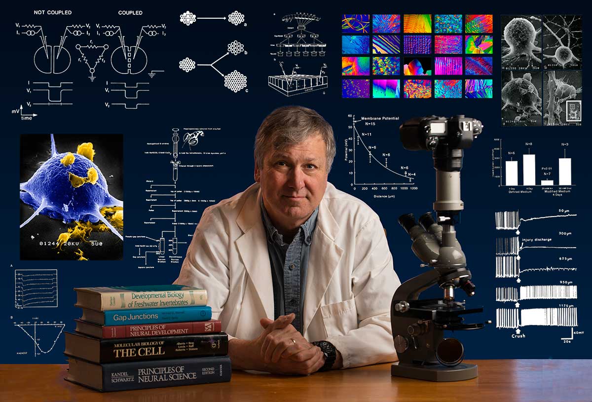
Bio: Robert Berdan is a professional nature photographer living in Calgary, AB specializing in nature, wildlife and science photography. Robert retired from Cell\Neurobiology research to pursue photography full time many years ago. Robert offers photo guiding and private instruction in all aspects of nature photography, Adobe Photoshop training, photomicrography and macro-photography. Portrait of Robert by Dr. Sharif Galal showing some examples of Robert's science research in the background.
Email at: rberdan@scienceandart.org
Web site: www.canadiannaturephotographer.com
Phone: MST 9am -7 pm (403) 247-2457.