
Photographing Microscopic Plant and Animal Life - Pond Scum II
by Dr. Robert Berdan
August 2, 2017

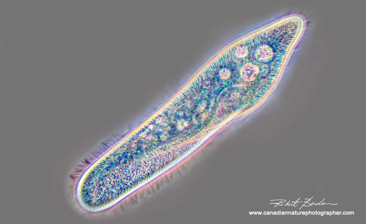
Stentor - a single celled Protozoan photographed with polarized light 400X The Stentor is dark but highly birefringent algae inside it appear yellow.
I am having so much fun exploring miniature universes filled with aliens - microscopic life - in water that this is my 6th article on photomicrography this year. I realize that not all nature photographers have a microscope, but those that are interested can purchase a used one from Kijjii and E-bay for only a few hundred dollars. Of course their are some expensive microscopes and I am saving for one with special optics called DIC (Differential interference optics) that will likely cost me $10,000 or more for a used one. But anyone can get into photomicrography and even use their cell phone to capture pictures. By using polarizing filters and a few accessories you will be able to take pictures like those shown below. Phase contrast is a more more expensive option as it shows the contents of living cells without staining them but it doesn't add colour to the subject like a polarizing microscope. Both techniques are valuable tools for studying microorganisms and cells in biology and in this article I use both techniques to take pictures of pond scum.
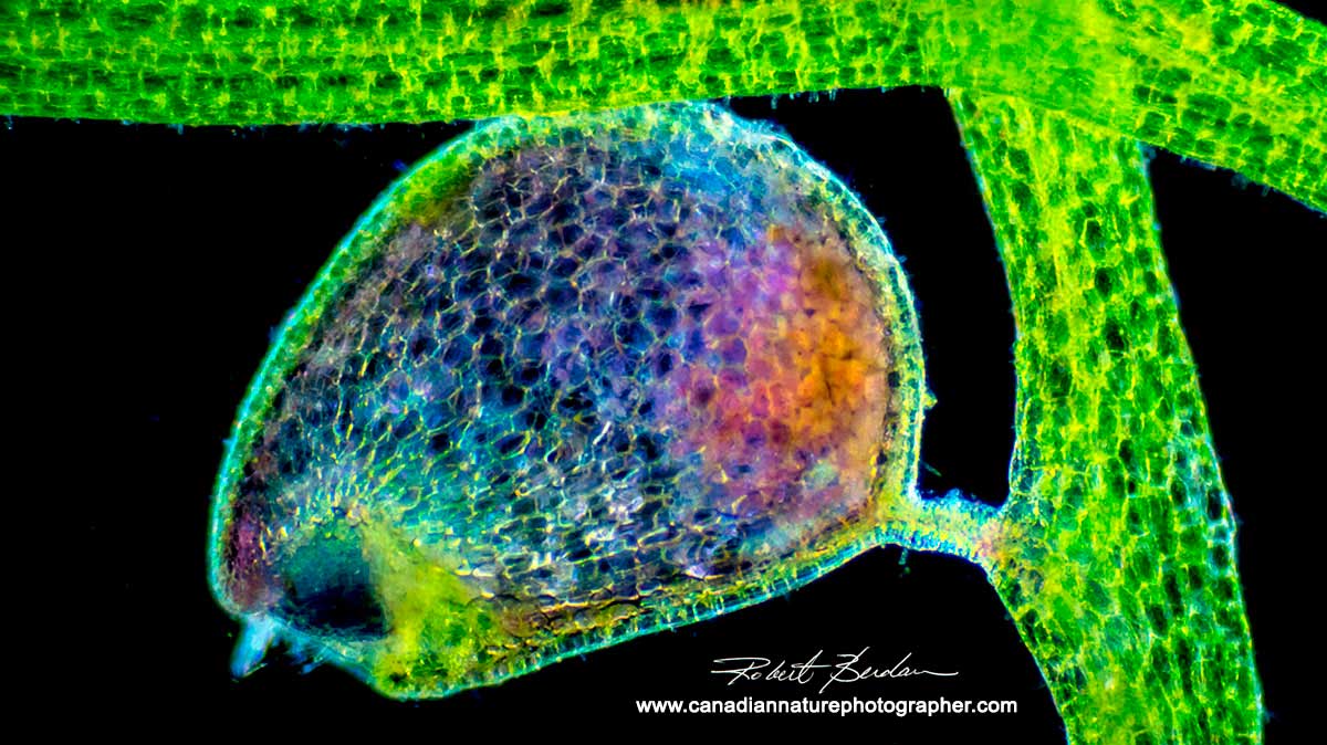
Bladderworts are carnivorous plants that live in lakes and ponds. Small crustaceans, rotifers and protozoa get trapped inside the bulb and are then absorbed by the plant. This bulb was photographed at low magnification using polarized light at 40X.
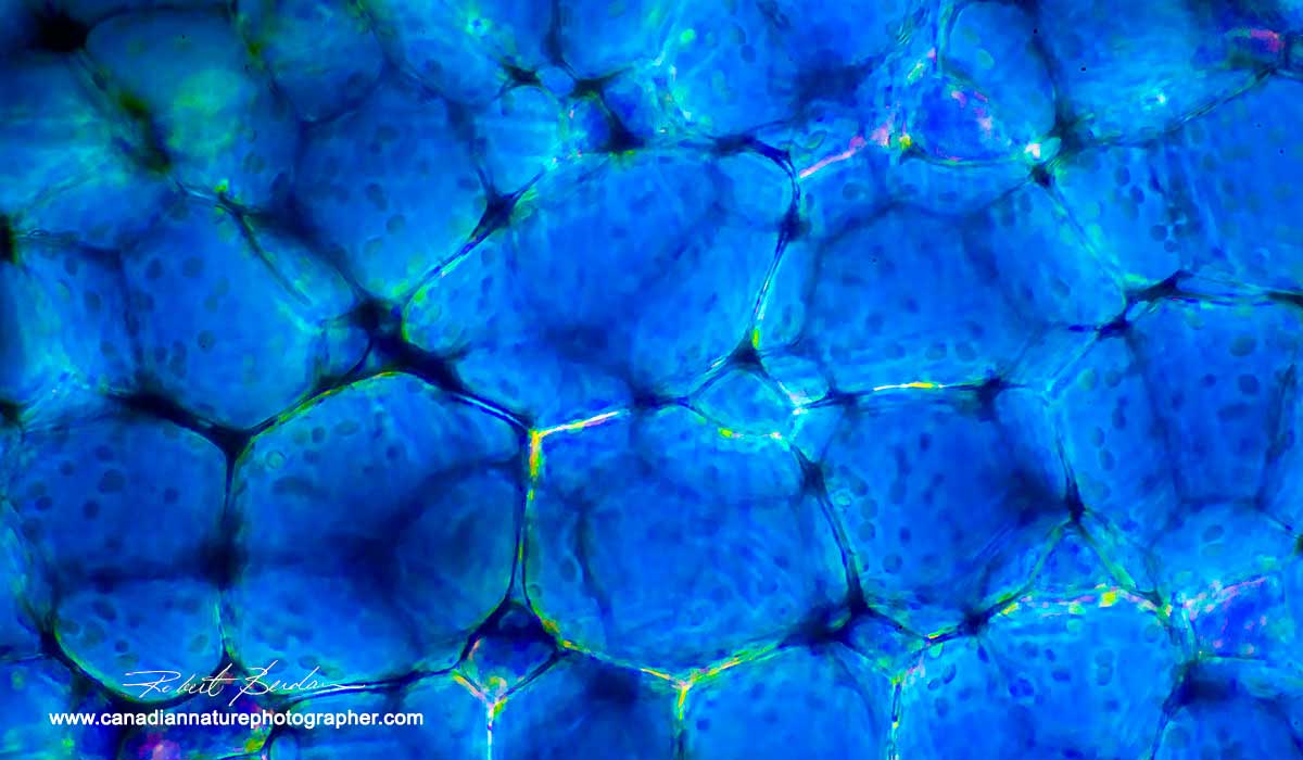
Bladderwort cells viewed in polarized light with a microscope. The dark small round objects are chloroplasts where photosynthesis occurs 100X.

The bladderwort plant has spines on some of its stems presumably to discourage fish and other predators from eating them. Polarized light microscopy and wave plate 100X.

Cells of bladderwort plant viewed in polarized light and a wave plate 100X.

Cladophora algae by polarized light microscopy 100 X

Cells within an aquatic plant showing the individual cells - 100X viewed with polarizing microscopy.
Adaptors to attach your camera to the microscope can be purchased for for under $100 or you can even make your own using a photographic extension tube and pop can or plastic tubing held together with electrician tape. Taking pictures through a microscope does involve an investment in time to learn how to do it. You will also have the satisfaction of knowing that fewer photographers are using a microscope. It's also easier then ever using a digital camera as your practice photos are free, you have instant preview and the camera can be coupled to a laptop using free software called Digicam control. There are also dozens of YouTube videos that will show you how to connect your camera or cell phone to a microscope (e.g. YouTube). The main problems in photographing through the microscope are 1) need for a very bright light source 2) high shutter speed to stop action of many microorganisms 3) vibration of the camera shutter can result in unsharp pictures 4) narrow depth of field especially at higher the magnification. Focus stacking helps improve the depth of field, but it requires that you capture several pictures at different focus points when the animal is not moving. To learn more about focus stacking see this article.

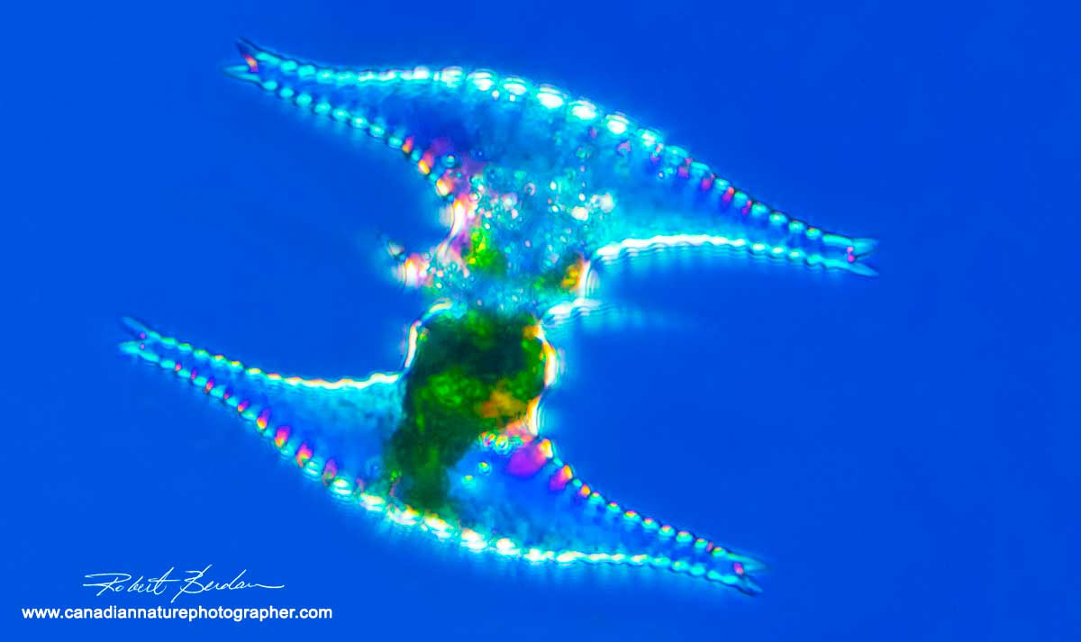
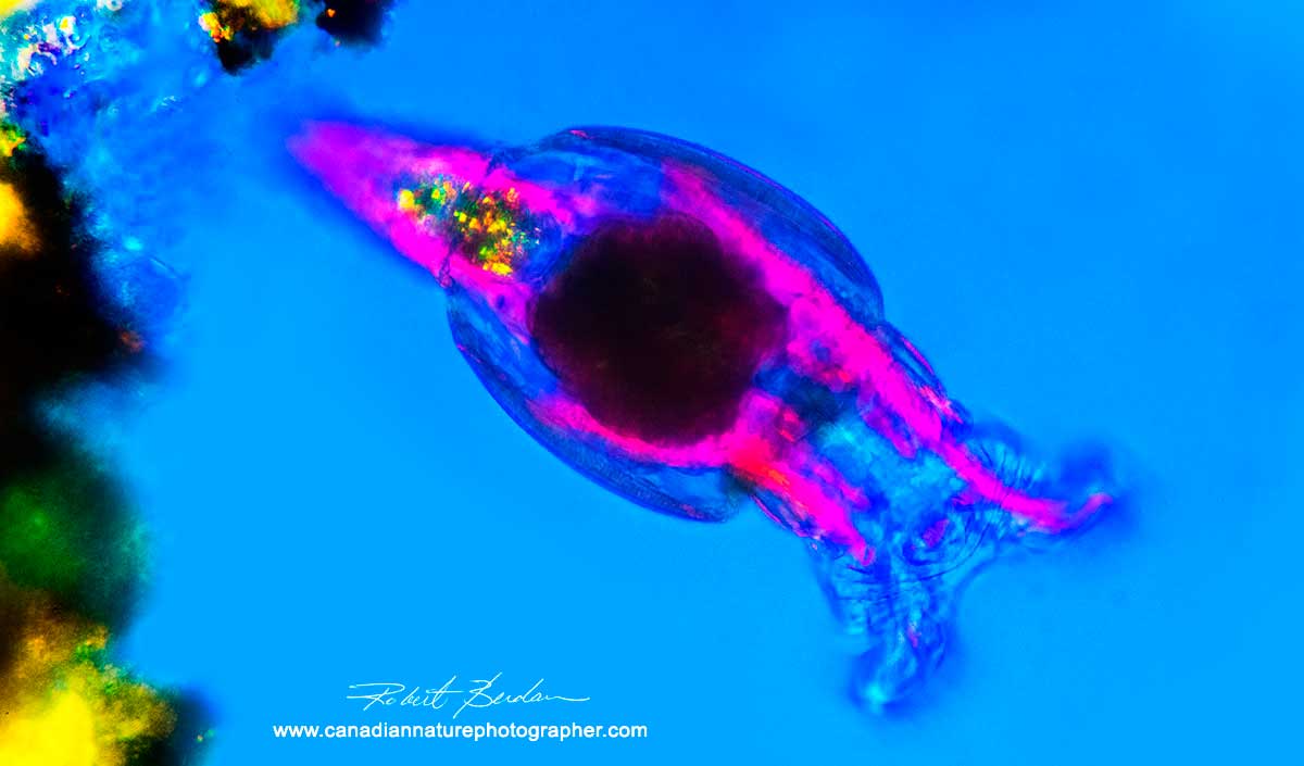
Dinoflagellate - Ceratium hirundinella are single celled protozoa that have hard shelled plates (theca made of cellulose ), two horns and they move through the water using flagella. Found in both salt and fresh water - I found this specimen in water from Twin Lakes, near Rocky Mountain house, AB. 400X. Polarized light microscopy, focus stack of 8 images.

Ceratium hirundinella. 400 X bright-field, phase-contrast, and polarizing microscope views - all focus stacked. Generally these dinoflagellates are harmless to humans, but under certain conditions can grow rapidly to produce a bloom known as a red tide that can deplete the dissolved oxygen in the water and kill fish. To learn more - see Wikipedia.
Desmid Staurastrum sp - single celled algae that forms birefringent shells that can appear jewel like as shown in this photo-micrograph taken with a polarizing microscope 400X.
The recent article "The Hidden World - My Look at Photomicrography by Marek Miś" using a microscope with polarized light to capture Desmids (single celled algae) inspired me to look harder for some of these organisms. I drove to Kananaskis via Sibbald Creek Trail and also drove to Twin Lakes near Rocky Mountain house in search of Desmids and other cool microorganisms. I always bring my DSLR (digital single lens reflex) camera in case I encounter wildlife or wildflowers and so I can photograph the ponds and streams where I collect water samples.
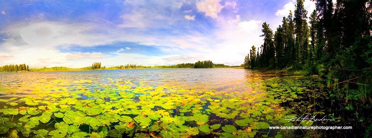
Above is a panorama of Twin Lakes with its numerous lilly pads surrounded by Boreal forest.
If you have access to a stero microscope you can collect some of the large insect larvae or use a light with a magnifying glass to reveal some of the larger subjects such as insect larvae (I found a light with magnifying glass at Canadian Tire for under $50). Anyone can start by examining pond water collected into jars with a simple magnifying lens and seeing if they can identify some of the larger macroscopic life and algae.

Cladophora a common water weed, viewed with Polarized light microscopy 100X
I own two light microscopes, a Nikon Optiphot which I use primarily for polarized light studies and my 45 year old Olympus E-microscope with positive low phase contrast. These techniques allow me to see translucent microorganisms and plants living in the water. For collecting specimens I simply take a few jars. I fitted my insect collecting net with a small plastic vial on the end and dip it through pond water to concentrate the organisms. I also bring a small aquarium net available at pet stores. For desmids I sometimes squeeze the cap of the jar down into wet moss and allow the water to collect and then pour the water into the jar. All this makes me feel like a kid again exploring the natural world. My wife often assists me and sometimes gets "soakers" walking in the wet moss around lakes and ponds. I also collect specimens (flatworms) from the underside of stones in creeks and other quiet pools. Sometimes I find "biological gold" an amazing assortment of creatures that you can see in my pictures below. I own lots of algae and protozoa field guides to help me identify the organisms. The incredible variety of creatures sometimes makes identification difficult unless one is an expert - nevertheless I try working through the keys and refer to the many diagrams. Some are easy, others I have not been able to identify for certain.
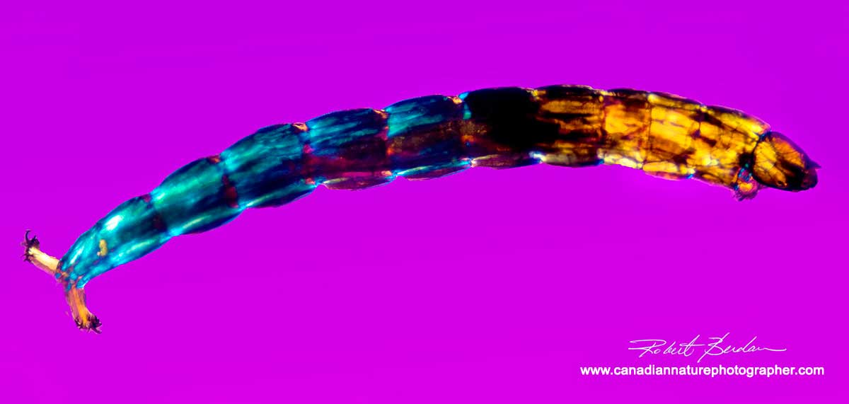
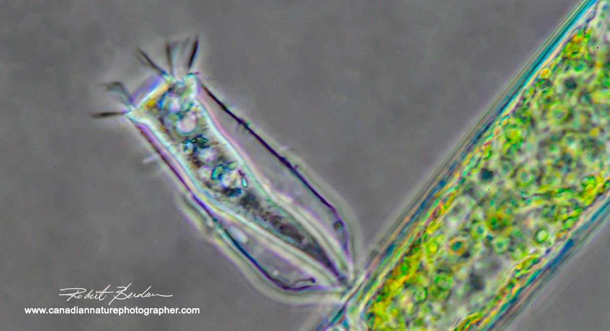
Chironomid - panoramic stitch of 3 images 40X polarized light microscopy. The Chironomidae are a family of midge flies that resemble mosquitoes. The larvae are quite common in pond water and can be seen by eye as they can grow to a cm or more. You can pick them out simply by looking at your jar of collected water as they wriggle. Mosquitoe larvae look similar - watch this YouTube video to see what they look.
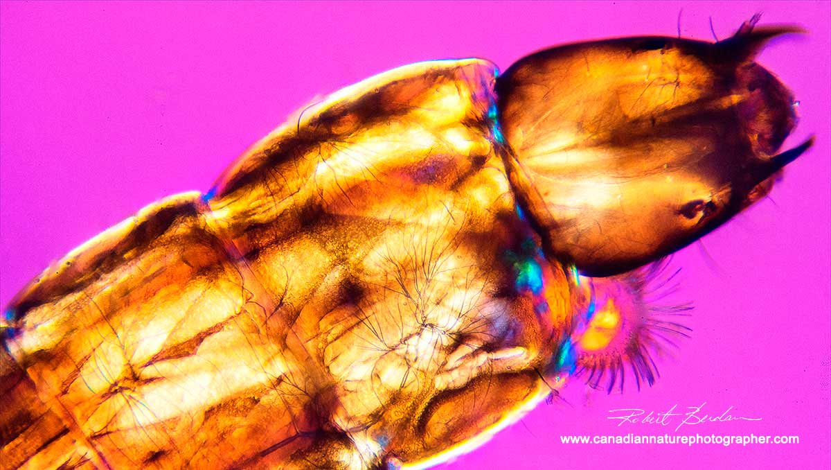
Closeup of the head of a chironomid you can see the trachea (small dark branches below the cuticle) the insect uses to distribute oxygen. Muscles are birefringent and show up bright yellow in polarized light. 200X polarized light microscopy. The pink background is created by inserting a wave plate between the polarizers (you can make a wave plate using scotch tape on glass or sometimes place a clear plastic CD cover between the polarizers and strain birefringence will produce various coloured backgrounds). Depending on how the wave plate is turned between polarized light you can get different coloured backgrounds - pretty cool. Note polarizing filters will only light up subjects that are birefringent with molecules organized like a crystal e.g. muscle, cuticle, cellulose, hair etc.

Posterior part of the Chironomid has hooks 200X polarized light microscopy

Gammarus is a common freshwater amphipod that can be easily seen by eye as they be several millimetres across. Photographed with Rheinberg filter. 20X.
When I observe these simple organisms, many of them made up of a single cell or small number of cells, it's amazing how complex many of their behaviours are and how well suited they are to their environment. Some of them, especially those that live in ponds that dry up, can form cysts that allow them to survive without water for months or years in a state of dormancy with virtually no metabolism. Ideal properties for beings if they plan to travel though space one day. Some of the algae (e.g. Blue greens) are thought to have been around the planet 3-4 billion years old according to fossil records. This has made me think about how life first began. We don't know the answer to this question but the minimal Genome project has found that there are about 360 genes necessary for life. Scientists have created a genome using a computer and put it into yeast cells that had their genomic machinery removed and these cells grow and reproduce. It seems likely that within the next decade that scientists may be able to create a single living cell from scratch. For those interested in how early life may have begun I recommend an article by the BBC.
Blue-Green algae sometimes called Cyanobacteria are one of the most primitive organisms living on the planet. There are basically three types of cells procaryotes, eucaryotes and archean cells. The procaryotes include bacteria and blue-green algae. They are the simplest cells on earth, they have no nuclear membrane, the DNA floats in the cytoplasm, they have no organelles like mitochondria or chloroplasts. Fossil evidences suggest that they fist appeared on earth between 3-4 billion years ago, the earth is estimated to be 4.7 billion years. The evidence for the oldest fossils comes from right here in Canada. Stromatolites another structure created by blue-green algae have been dated to several billion years old. Fossils of blue-green algae that I have looked at are very similar to the photos I show below and it makes me ponder.

Anabaena filament showing the individual cells, some of the cells are specialized to fix nitrogen (Heterocysts - 2nd from left) and other Akinetes (spore cells - left tip). Some blue-green algae can produce neurotoxins that during a bloom can kill water fowl and cattle. 500X phase contrast microscopy.

Merismopedia is another blue green algae that forms in regular flat packets embedded in a mucilaginous matrix 500X

Aphanocapsa blue-green algae that also forms in irregular mucilaginous matrix 500X
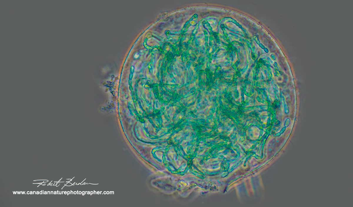
Blue green filamentous algae (Nostoc sp) that formed inside a tight ball 200X Phase contrast microscopy.
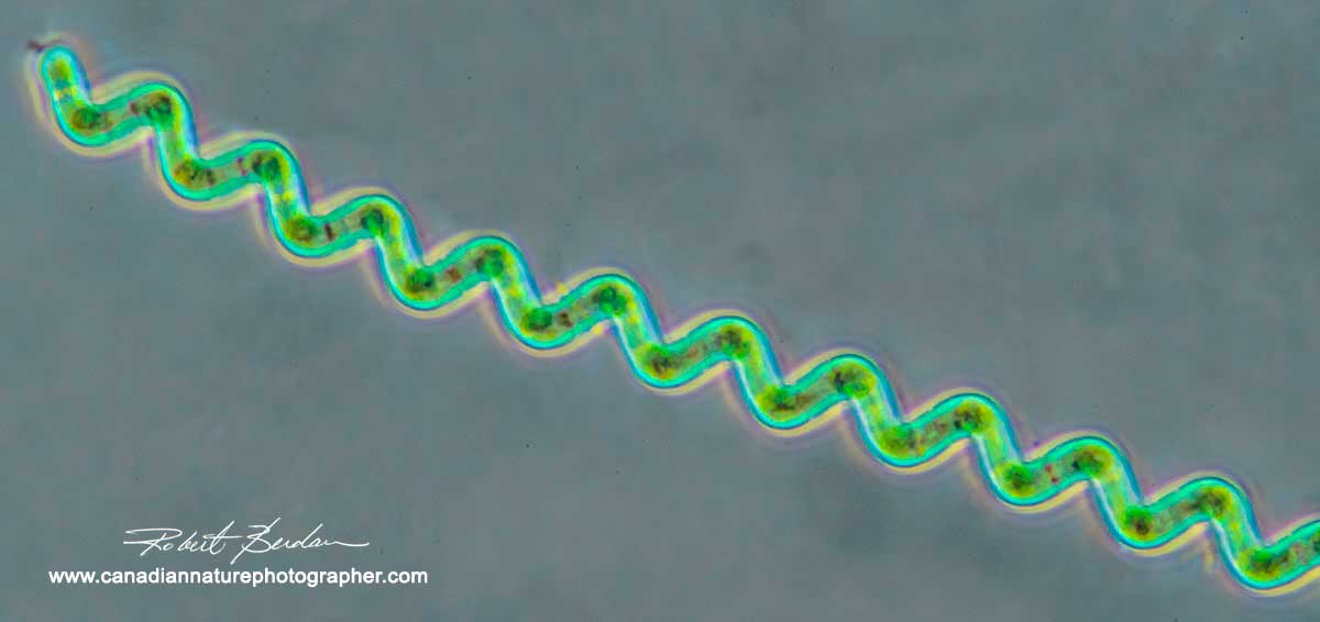
Spirulina is a blue-green algae that appears to move through the water in a spiral fashion. 400X Phase contrast microscopy. Some species of this group are cultivated and used as a dietary supplement available in some health food stores and are used to fertilize rice paddies.
Rotifers are sometimes called wheel animals because many of them have cilia that appear to beat in circles near the head. The cilia move bacteria into their mouth and also provide locomotion. There are thousands of species of rotifers that range in size from about 0.1 mm (100 microns) to over 2 mm (2000 microns) in size. They can form cysts that can withstand dessication and live in suspended animation for decades without water. They are common and often found near or attached to water plants. They are some of the most interesting animals I have observed with the microscope. They are composed of about 1000 cells, some include eyes, a primitive nervous system and 99% are females, males have only one purpose to fertilize eggs and don't even have mouth parts. Rotifers are also used in studies of aging (for Geeks only).

Bdelloid rotifer viewed in Phase contrast microscopy, you can see the rotary wheels at the right side of the picture covered in cilia with a distinct space separating the ciliated wheels. It is fascinating to watch rotifers swim and crawl around 200X.
Bdelloid rotifer in polarized light. The polarized light has made the muscles pink in this picture. 200X

Rotifer in polarized light - by changing the rotation of the polarizers and a full wave plate I can produce different colours in the parts of the animal that are birefringent (have crystalline properties that rotate polarized light).

Rotifer (Monostyla sp) with a red "eye" Phase contrast microscopy 400X
Desmids are unicellular green algae that often have bilateral symmetry and crystalline properties when viewed by polarized light microscopy. They are found in lakes, ponds and sphagnum moss in marshes and often favour water with a low pH (4.0) which is where I have been looking for them. Along with the Diatoms they are the true jewels of the microscopic world.

Cosmarium sp. belongs to the group of green algae called Desmids. Viewed under polarized light microscopy 400X

Staurastrum sp is a unicellular green alga belonging to the order Desmidiales. These jewels look like "alien space ships" under polarized light microscopy 400X.

Pleurotaenium sp Desmid - in polarized light 400X with a full wave plate which produces blue background.

Spinocosmarium sp Desmid in polarized light microscopy 400X

Cosmarium Desmids Polarized light microscopy 400X

Micrasterias Desmids viewed by phase contrast microscopy 300X.
Protozoa - there are thousands of species, the most familiar are the amoeba and paramecium which I captured below using phase contrast microscopy. The main challenge with paramecium and other ciliated organisms is slowing them down. This can be done by adding methyl cellulose to the water, heating the slide gently for about 5 seconds on a hot plate. To stop the cilia motion I am working on using my flash and will present an article in the future once I master this technique in the microscope. My problem is when I put the flash under the scope I can't see what I am photographing - other microscopic e.g. Charles Krebs has built his own light source with a built in flash.
Paramecium by phase contrast microscopy 400X. Note the numerous cilia (hairs) that surrounds this one celled organism. The cilia enable them to move very fast. I was able to subdue this specimen by drawing the cover glass down by draining the water using capillary action produced by the edge of a paper towel. In early days microscopists used special coverslip compressor e.g. a Rousselet compressor or Taylor compressor.

Amoeba sp via phase contrast microscopy. These one celled animal moves more slowly and are easier to photograph. The amoeba are fascinating to watch as they crawl by streaming their pseudopodia (false feet). 400X .
Ciliated Protozoan Thuricola foliculata that lives in a lorica attached to algae 400X Phase contrast microscopy. When the animal is touched by another organism it rapidly contracts to the safety of the lorica. A lorica is a shell-like protective outer covering, often reinforced with sand grains and other particles that some protozoans and loriciferan animals secrete. Usually it is tubular or conical in shape, with a loose case that is closed at one end.
Diatoms (Didymosphenia geminata ) growing on a stalk attached to detritus, 400X Phase contrast microscopy.

Diatom Navicula sp. 400X Phase contrast microscopy. They have an outer silica shell and internal brown photosynthetic pigments and oil droplets. They are common in fresh water and soil and glide slowly on the microscope slide.
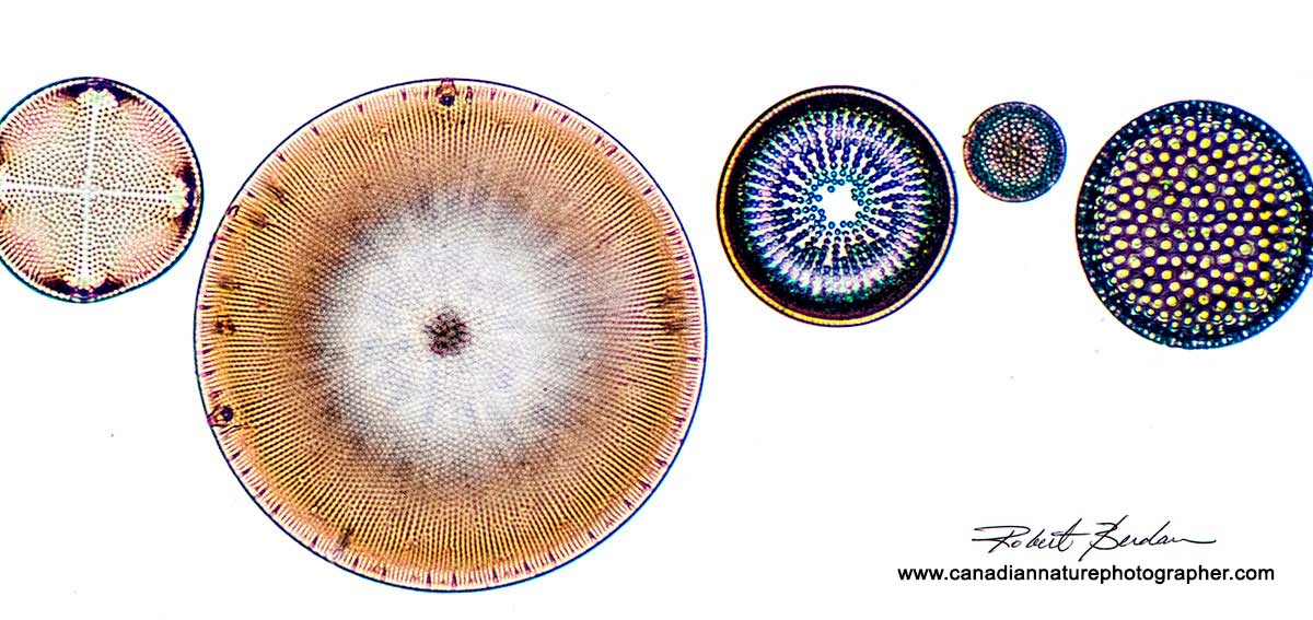

Fresh water diatoms are single celled animals that form silica (glass) shells in a wide variety of patterns. They are one of the most common organisms on the planet and you will find them in almost any pond or stream. Many are on the bottom or cling to aquatic plants. The top two pictures are from prepared slides of Diatoms photographed by bright field microscopy 400X.

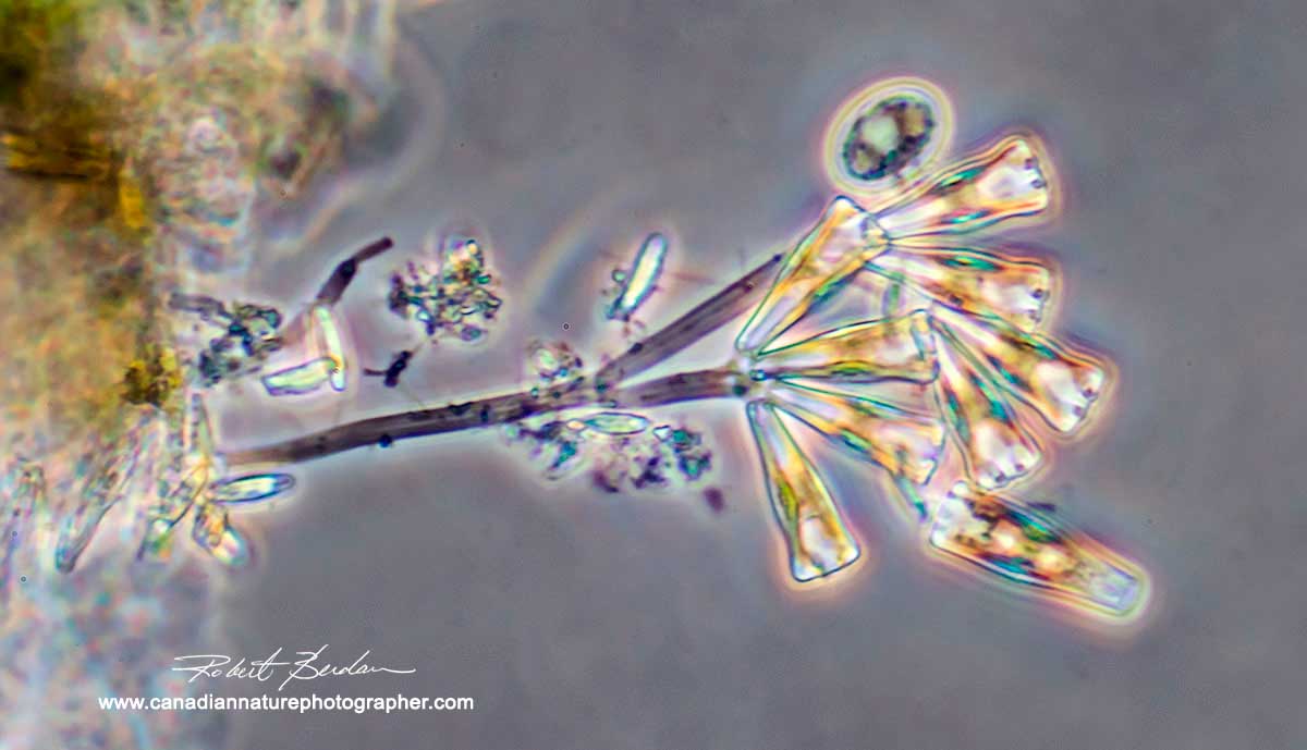
Mix of Desmids, diatoms, and protozoa 400 X phase contrast microscopy. a) Netrium sp Desmid b) Spinocosmarium sp. Desmid c) Fragilaria sp Diatom d) Gomphenama sp. Diatom e) Pediastrum Boryanum algae f) Dinbyron lorica flagellated protozoan g ) unknown - crustacean larvae ? h) Lacrymaria sp ciliated protozoan i) ciliated protozoan unknown species
Note in some of my photographs I cleaned up the backgrounds by removing dirt, out of focus highlights, and bacteria that were a distraction, the biological integrity of the organisms was never compromised in any photos.

The microscopes I used to take the pictures in this article include on the Left: Nikon Optiphot I used for polarizing photos equipped with Nikon D800 , on the Right: Olympus E-microscope used for Phase contrast microscopy equipped with a Nikon D500 - I have owned this microscope for 45 years. On the left I am using an AM Microscope Camera adapter CA-NIK SLR to attach my Nikon D800 and on the right I am using a Nikon extension tube attached to an Olympus Microscope Photo tube using black electrician tape to attach my Nikon D500. I am using 5X and 3.3X NFK photo eyepieces. Both microscopes are connected electronically to my Laptop computer using USB cords and the images are caputured to disk using Digicam control software (free). I upgraded my Nikon Optiphot light source recently to a 30Watt LED lamp from E-bay. LEDs light sources are available for other older microscopes and I recommend them. Note for my Olympus microscope I am using a Kodak slide projector to provide a bright light source. In order to get faster shutter speeds I often set my ISO speed to 800-1600 and record RAW files. I reduce digital noise in the RAW files using Adobe Photoshop Camera RAW.
If you might be interested in learning photomicrography I can offer you instruction and advice on buying a scope. Microscopy first led me into a career in Science and later to become a nature photographer. I have invited more microscopists to show their pictures and share their techniques in the future and I hope this might inspire some of you to try photomicrography. RB
References and Additional Links.
Digicam control software for coupling your camera attached to a microscope to laptop - Free for Windows.
AM microscope - photography adapters for attaching your camera to a microscope
Microbe Hunter - beginning guide to microscopy web site - good resource
Starting with Microscopes - The Quekett Microscopical Club
How to use a Microscope and Take a Photomicrograph - Nikon PDF
Take Digital Photos Through a Microscope Without Any Special Lens
Charles Krebs - photomicrographer includes many good articles on how he takes photomicrographs
Nikon small world 2016 Photomicrography Competition view some of the best photomicrographs.
World in Motion - videos taken with light microscope
The idea of Life Began as Crystals - BBC
The secret of how life began - up to date research - BBC
Earliest evidence of Life - BBC
Oldest evidence of life 3.8 billion years old
Desmids - Wikipedia
Cyanobateria - Blue green algae Wikipeadia
Algae Identification field guide - Canadian Government - PDF
Wave plate - Olympus microscope - for Geeks
Custom built LEDs for older microscopes
- E Bay
Also see my other articles on Microscopy:
Scanning Electron Microscopy - Photography
Rheinberg filters for Photomicrography
The Art & Science of Photomicrography with Polarized Light
Microscopic Life in Ponds and Rainwater
Photographing Through a Microscope Photomicrography - Inner Space
Using a scanner as a low power light microscope
Authors Biography & Contact Information

Robert Berdan is a professional nature photographer living in Calgary, AB specializing in nature, wildlife and science photography. Robert offers photo guiding and private instruction in all aspects of nature photography and Adobe Photoshop training.
Email at: rberdan@scienceandart.org
Web site: www.canadiannaturephotographer.com
Phone: MST 9am -7 pm (403) 247-2457.
Click on the buttons below and share this site with your friends