
Curved Crystals viewed by Polarized Light Microscopy
Amino acids, Vitamin C, Menthol & Caffeine
by Dr. Robert Berdan
January 2, 2021

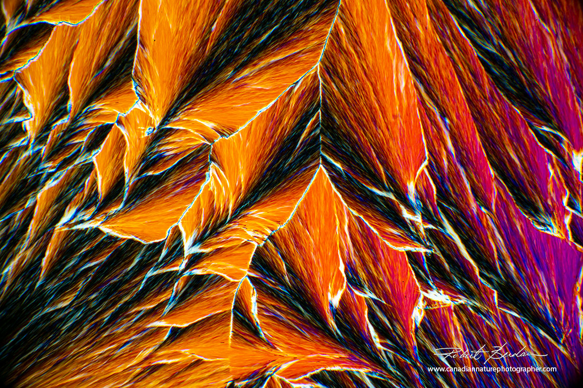
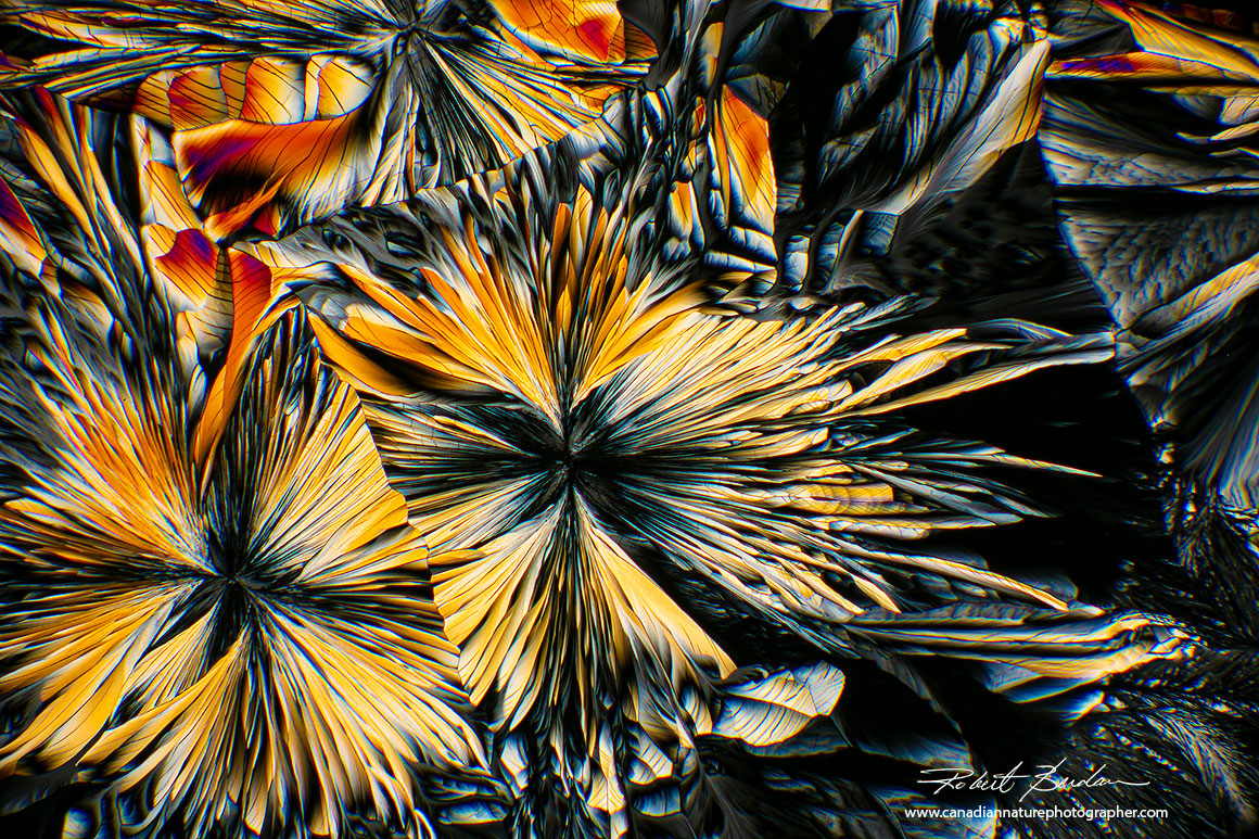
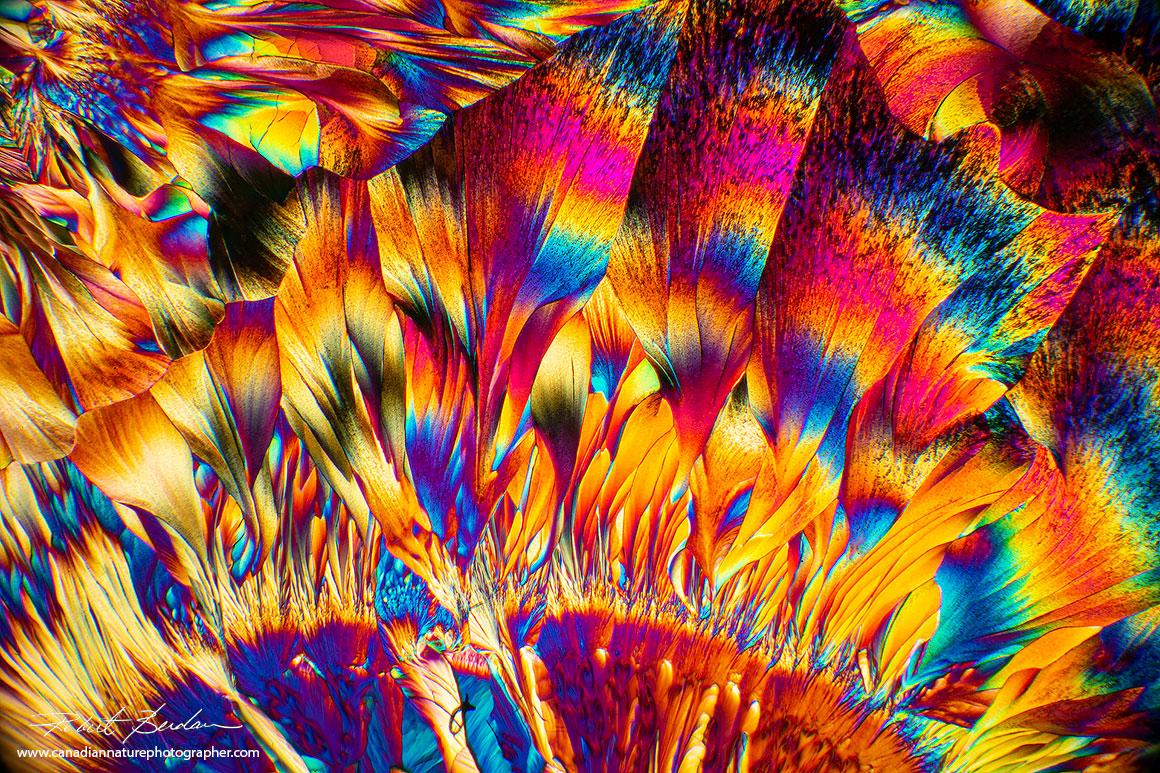
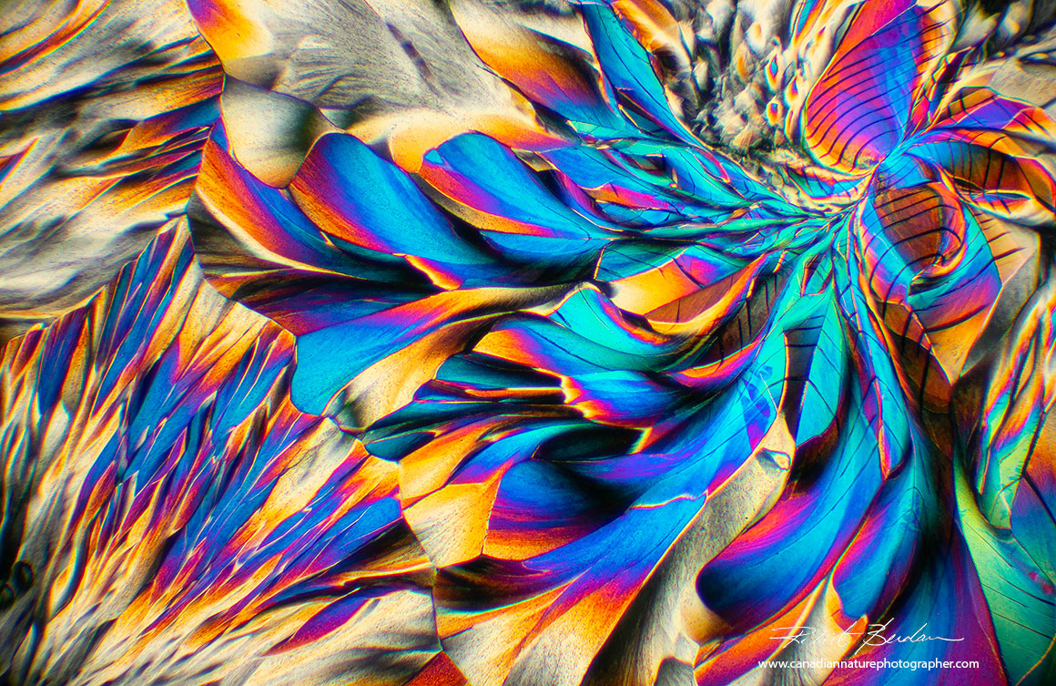
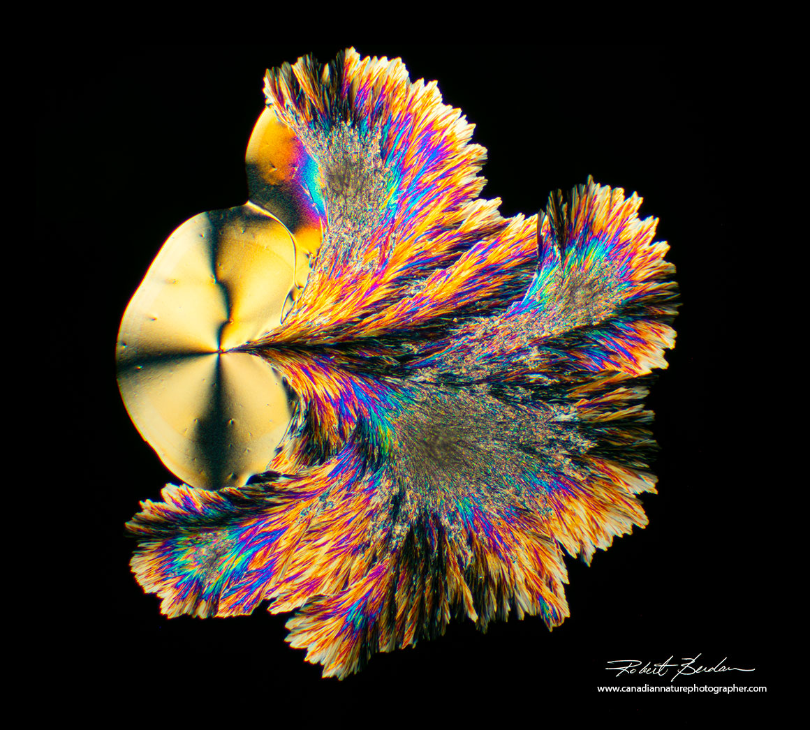
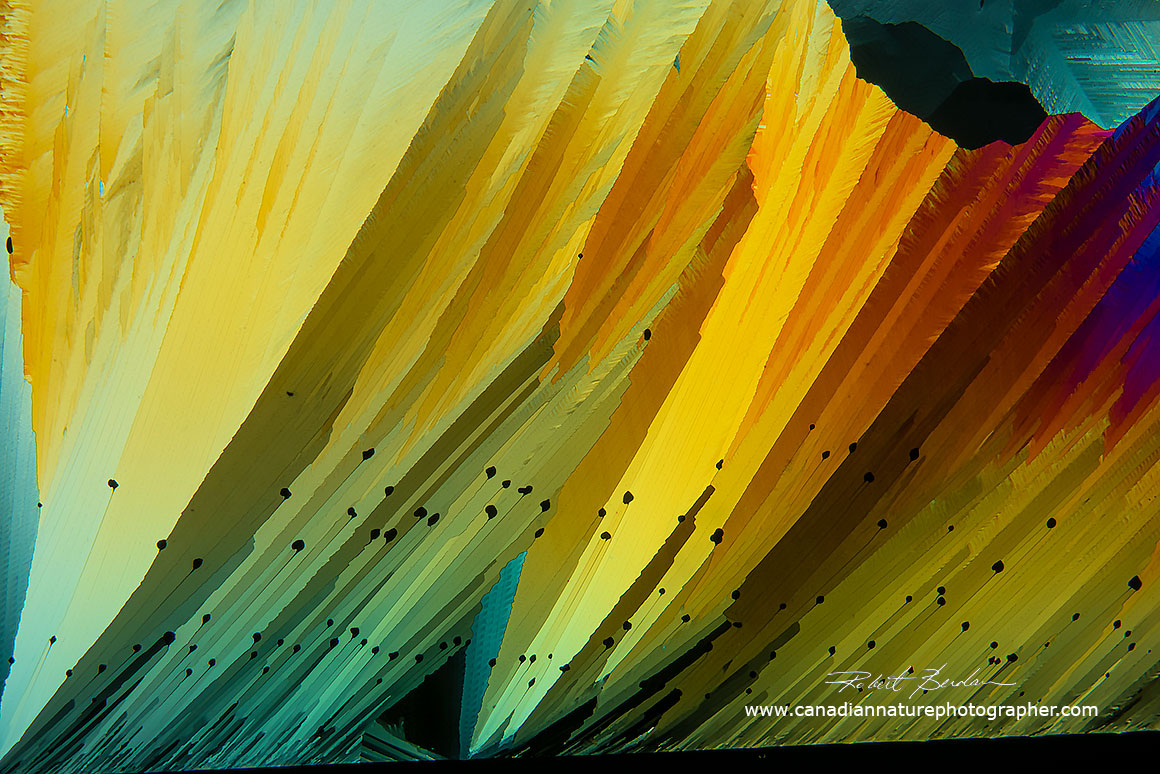
Crystals of Beta-Alanine and Glutamine produce flower-like crystals when dissolved in 1 part 95% ethanol (Everclear 190 proof) and 1 part deionized water and then warmed on a microscope slide -100X via polarizing microscope. Some images resemble abstract paintings.
Introduction
Microscopic crystals can exhibit an array of colours and shapes more spectacular then just about anything else I have seen in nature. The colours for the most part are produced by light interference and can exhibit rainbow colours. To view these colours is simple if you own a light microscope with a pair of polarizing filters. One polarizer is placed below the slide often on the light source or bottom of the condenser and one above the objectives usually in the microscope binocular housing or eyepieces. The polarizers are rotated to extinction position which blocks the light coming through and produces a black background. Certain anisotropic crystals twist the light rays (basically anistropic crystals separate the light rays that travel at different velocities through the crystals - see birefringence) and colours can be seen when viewed through crossed polarizers due to interference. At the end of the article I provide links to movies and articles on how to convert a microscope to use polarized light.
You can purchase a microscope specialized for viewing specimens in polarized light but they are not necessary to view crystals like that shown in this article. A proper polarizing microscope offers a rotating stage, the ability to insert compensating filters also called wave plates (full wave, 1\4 wave etc) that can modify and enhance the interference patterns. Cheap wave plates can be made from Scotch brand transparent tape, thin sections of mica or using a piece of plastic from a plastic compact disc box. Some polarizing microscopes offer a Bertrand lens which allows you to see (conoscopic) uniaxial and biaxial interference patterns of crystals. For information about a polarizing microscopes see my recent review of the Motic BA310 microscope which I purchased a few months ago and used it to create the pictures in this article. Note most of my photomicrographs of crystals were taken using low power objectives 4, 10 or 20X. If I needed a wider field of view I made panoramas by stitching between 2 to 40 or more overlapping images in Photoshop.

Vitamin C crystals by Polarized light microscopy 40X. Note the bright coloured interference colours. Motic BA310 polarizing microscope.
Crystals that appear coloured in a polarizing microscope are said to be birefringent, that is they split the polarized light into 2 beams that travel through the crystal at different velocities and then interfere when they leave the crystal. Interference causes certain colours to be eliminated from white light due to destructive interference and other colours to become brighter due to constructive interference. At the end of the article I provide links to more information on how this process works. What is important to know is that that many biological substance are also birefringent e.g. hair, wood, starch grains, bone etc - almost anything where the molecules are arranged in an organized crystalline pattern may be birefringent. For a more detailed explanation of birefringence and anistropy with polarized light see Lordana M. Völker-Pop (2014) - PDF.
Menthol crystals formed using the melt method 40X polarized light microscopy.
Most crystals I prodced have straight edges like that shown below. Some crystals however form round shapes (e.g. Vitamin C) and some amino acids can form wave-like shapes that can resemble flowers and leaves. Crystals with curved lines are found less often. In this article I will present the methods I use to achieve curved crystals. I believe they are some of the most beautiful patterns that can be observed with a microscope and each crystal is unique.

Many crystals form sharp rectangular arrays and points with bright colours - Saccharin 40X

Under some conditions Saccharin (a sweetener or sugar substitute) when viewed by polarized light microscopy 100X will form flower like shapes. Small changes in the crystallization conditions can produce vastly different shaped crystals.
Basic Methods of Making Crystals for Viewing by Polarized Light Microscopy
There are two main methods I use to make crystals for viewing by polarized light microscopy. The first method is called the Melt method and it requires a hot plate or Bunsen burner (some folks use a microwave). You obtain some pure chemicals from a pharmacy, nutrition store, medicine shop, or Amazon e.g. caffeine, Vitamin C. Then look up the melting point of the crystals - the lower the melting point the easier it is to produce a melt which are crystals that form upon cooling after melting them. Caffeine and Citric acid are good chemicals to start with. Note that some chemicals won't melt, they may turn brown and burn and some chemicals can give off toxic fumes (e.g. Benzoic acid) so do some research before you experiment with any chemical.
Also you can melt the chemical on the slide alone, or I sometimes cover the chemical with a coverslip so the crystals form in an even flat sheet upon cooling e.g. caffeine. I recommend you start with chemicals having low melting points i.e. below 300°C - glass has a melting point of 1700°C. Place the slide and chemical on a hot plate and warm up the slide. Many chemicals will turn into a clear liquid when melted. Remove the slide with tweezers or forceps and allow the slide to cool. I usually place the slide on a porcelain bathroom tile (available for about $1 at hardware stores). You can also place the slide on the microscope stage and watch the chemicals crystallize (see movie below) and take video of the crystals growing with your camera. Some crystals form quickly within a minute while others may require time-lapse photography (Digicam control software which is free can do this).

Vitamin C "microscape" - Vitamin C crystals dissolve readily in water and when placed on a slide may begin to crystallize within a few minutes. Vitamin C produces circular crystals and a variety of other interesting shapes. Adding a little bit of alcohol accelerates the dryng and crystallization and reduces the surface tension (some folks wash their slides with soap to reduce surface tension of water).

Vitamin C crystals forming circular discs upon cooling Polarized light microscopy 40X
The second method I use to make crystals is simple, make a concentrated solution of the chemical - I use plastic test tubes and a vortex mixer to make the solutions. I sometimes warm the solution in a water bath to increase the solubility of the substance. Then using a pipette I place several drops and\or smear the solution on a microscope slide, label the slide and then allow it to dry. You can accelerate the evaporation of the solvent by placing it on a hot plate at low temp. Sometimes the crystals get larger if you allow the solution to evaporate slowly for a few hours or overnight - you need to experiment to find the optimum conditions. To get started try some Epsom salt or Vitamin C in water or ethanol and place the solution on a slide. Epsom salts will crystallize very quickly. Sometimes the solution beads up on the slide due to surface tension of water. The surface tension can be reduced by adding a very small amount of alcohol to it. The solvent used to dissolve your chemical is important and it depends on the properties of the substance - some substances are more soluble in water and some are more soluble in alcohol and others require a combination of solvents (do a bit of research and experimentation).

Beta-Alanine and Glutamine mix in water\ethanol 40X Polarized light microscopy.
My recommendation for a solvent is to start with deionized water, if you don't have that use tap or bottled water. Also try isopropyl alcohol, methanol (toxic) and ethanol. Ethanol is available from some liquor stores (grain alcohol - one product Everclear 190 proof is 95% ethanol). Some folks use Vodka as a solvent (the higher the proof the more ethanol it contains e.g. 100 proof is 50%, 200 proof is 100% ethanol). You don't need a lot of solvent, 50-100 ml will be fine for hundreds of slides. Note that some substances are soluble in both water and alcohol or in a mixture of the two. The pH of a water based solution can be made more acidic by adding a weak acid like vinegar or more basic by adding sodium bicarbonate (baking soda). These are many variables you can try to promote the growth of crystals. Not all substance will form crystals. Many medicines e.g. aspirin pills may have substances added to them to inhibit crystallization. You can use a mortar and pestle to grind pills, or moth balls etc but some substances still may not form crystals, but the only way to be sure is try them and keep notes on what works and what doesn't. When you do this you are being an amateur scientist.
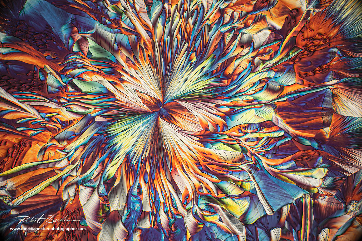
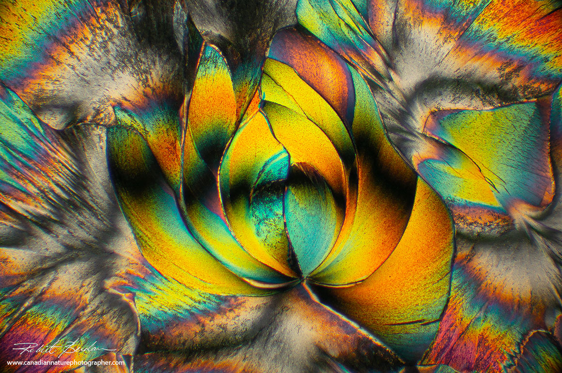
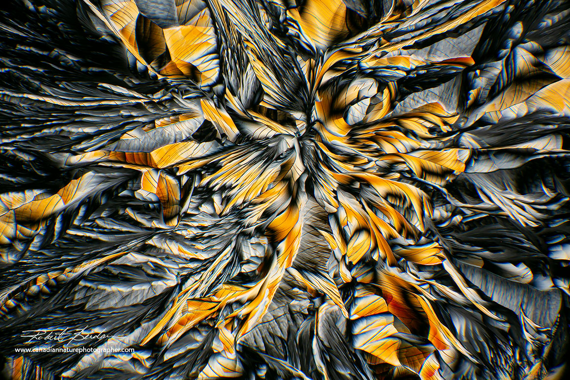
Flower-like image of Beta-alanine and Glutamine shows great complexity. In this image I also reduced the overall saturation using Photoshop.
Amino Acids for Making Crystals
There are 20 amino acids of which 9 are essential in our diet. Some amino acids are basic, acidic or neutral - learn more about their charge properties. Their charge determines their solubility in different solutions. I have only tested 4 amino acids: Beta-Alanine, L-Glutamine, Taurine and Lysine. Lysine did not form crystals for me after testing several solvents though it is sometimes used in research to make slides more adhesive to cells. I first tested each amino acid by itself in different solvents and only Taurine formed some crystals by itself. I tested them in water, 95% methanol, isopropyl and ethanol. I then tested Alanine and Glutamine in ethanol\water 1:1 and achieved the results you see in this article.
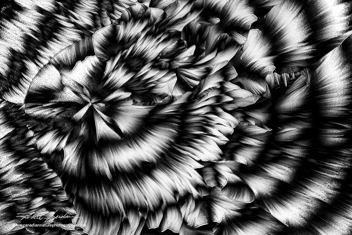
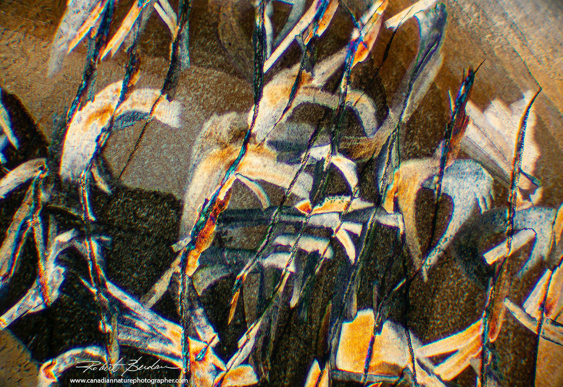
Crystals of Beta-Alanine and Glutamine sometimes formed black and white crystals in Polarized light. When the crystals are very thin they do not exhibit colour. Birefringence is dependent on the difference in the two refractive indices of the crystal and their overall thickness.

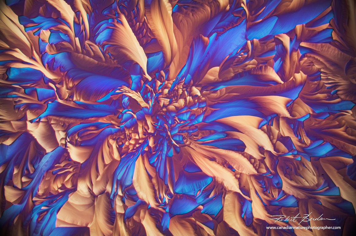
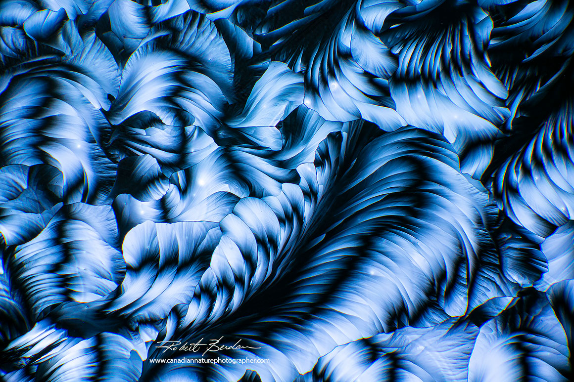
Amino acids Beta-Alanine and Glutamine formed a thin layer of crystals without colur. I added a blue tone to the images using Photoshop. Polarizing microscopy 40X.

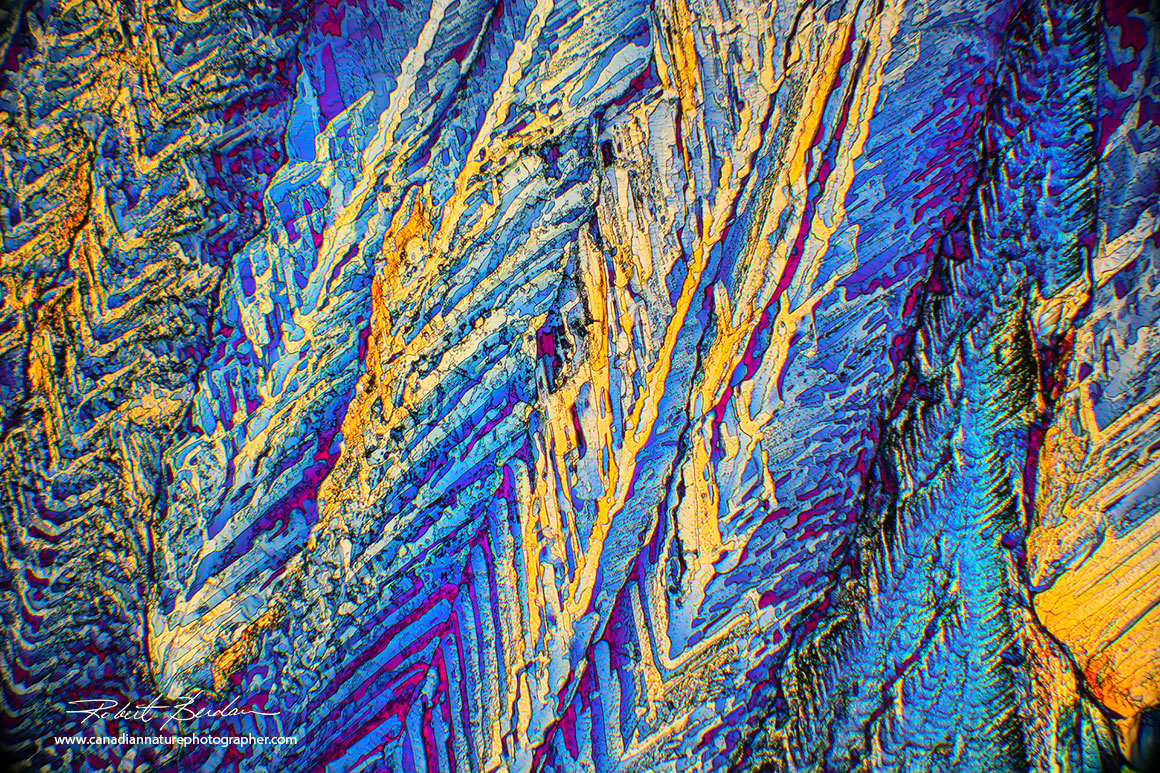
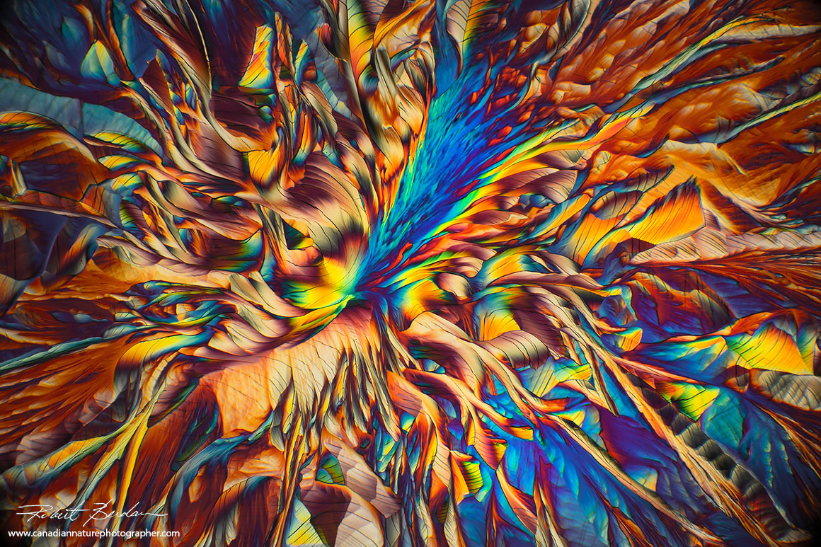
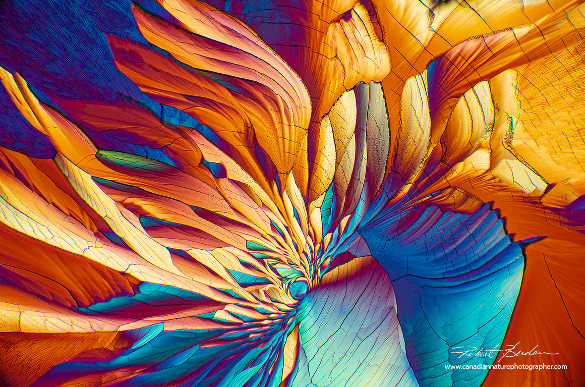
Sometimes the crystals of Beta-Alanine and Glutamine create crystals wtih intense colours that almost scream at you they are so saturated. 40X panorama.
Some microscopists use 40 proof Vodka as a solvent to make a solution of amino acids with good results. I tested 4 amino acids dissolved in water alone, then added vinegar or baking soda to alter pH - these solutions did not produce crystals except for Taurine in alcohol. When I used ethanol\water mix I began to see curved shaped crystals. I found that by heating the slides briefly (about 1 min on a hotplate) they would start to crystallize around the edges of the solution. I would then remove the slide to cool them briefly (30 sec to 1 min) then put them back on the hotplate for about a minute and they would begin to crystallize quickly. What seems to be happening is that the first heating creates small crystals around the edges of the solution that act as seeds that promote rapid crystallization after a brief second heating. You may need to experiment to achieve optimum results, but this procedure produced the best crystals in my hands. I have not experimented with different ratios of ethanol to water but it I believe its worth testing.

Note the more slides you create the luckier you will become. I recommend making at least 10 slides or more. I purchased my amino acids from a nutrition supplement store (Popeye's) in quantities greater then I will ever need. Amino acids are also available from Amazon - purchase the smallest amount you can as you don't need much. I recommend trying both Beta-Alanine and Glutamine mix in water\ethanol. I also made some slides with a mix of Beta-Alanine, Glutamine and Taurine that produced some interesting crystals (see below).
The amino acid Taurine dissolved in Ethanol by itself produced some crystals. 100X polarized light microscopy.

A mix of Beta-Alanine, Glutamine and Taurine in ethanol\water solvent dried on a microscope slide. Polarized light microscopy and a full wave (550 nm) compensator 100X.
Beta-Alanine and Glutamine 40X polarized light microscopy.
Beta-Alanine and Glutamine in ethanol\water 100X polarized light microscopy.
Beta-Alanine and Glutamine in water\ethanol 100X polarized light microscopy.
Beta-Alanine and Glutamine 100X polarized light microscopy. Image desaturated.

Beta-Alanine and Glutamine 100X polarized light microscopy.

Beta-Alanine and Glutamine 100X polarized light microscopy.

Beta-Alanine and Glutamine 100X polarized light microscopy.

Beta-Alanine and Glutamine 100X polarized light microscopy and full wave compensator.
Beta-Alanine and Glutamine 100X polarized light microscopy. This image made me think of the sun's corona with light emanating from the surface of the sun at the bottom of the picture.
Beta-Alanine and Glutamine 100X polarized light microscopy.
Beta-Alanine and Glutamine 100X polarized light microscopy showing crystals resembling flower petals.

Beta-Alanine and Glutamine 100X polarized light microscopy. Not all the crystals formed had curves or rounded shapes - there is a great deal of variability in crystal shape and form on some microscope slides.
Beta-Alanine and Glutamine 100X polarized light microscopy - toned blue in Photoshop.

Flower-like patterns. Beta-Alanine and Glutamine 40X polarized light microscopy.
Beta-Alanine and Glutamine 100X polarized light microscopy.
Beta-Alanine and Glutamine 100X polarized light microscopy, colours subdued in Photoshop

Beta-Alanine and Glutamine 100X polarized light microscopy.

Beta-Alanine and Glutamine 100X polarized light microscopy.
Movie of Crystals growing on a Microscope Slide
Movie made with Motic BA310 polarizing microscope and Canon 5D Mark III camera using Digicam Control software (free). Watch on YouTube https://youtu.be/GWESk2vMzMY
Miscellaneous Crystals by Polarized Light microscopy

Benzoic acid crystals form long linear crystals 40X polarized light microscopy. These crystals were produced using the melt method. CAUTION - Benzoic acid creates toxic fumes I suggest that you only try this chemical in a well ventilated area or outside as the fumes can affect your eyes and mucous membranes.
Sulfamic acid - the patterns resemble cave paintings or petroglyphs. Sulfamic acid is available at hardware stores and is used to clean concrete, ceramics, rust and limescale. This was a melt. Sulfamic acid can also produce cubic crystals and a variety of other shapes - also see my other article on crystals in polarized light. Polarized light microscopy 100X.
Vitamin C is readily available in stores, cheap and is highly soluble in water or alcohol. Simply apply a solution to a slide and wait until the crystals dry. Try water then alcohol (isopropyl, methanol or ethanol) or a mix as a solvent. A mix of water and alcohol seems to work best. Heating the slide accelerates crystallization, but the more slowly the crystals form the larger they tend to become - some can be several millimeters in diameter.
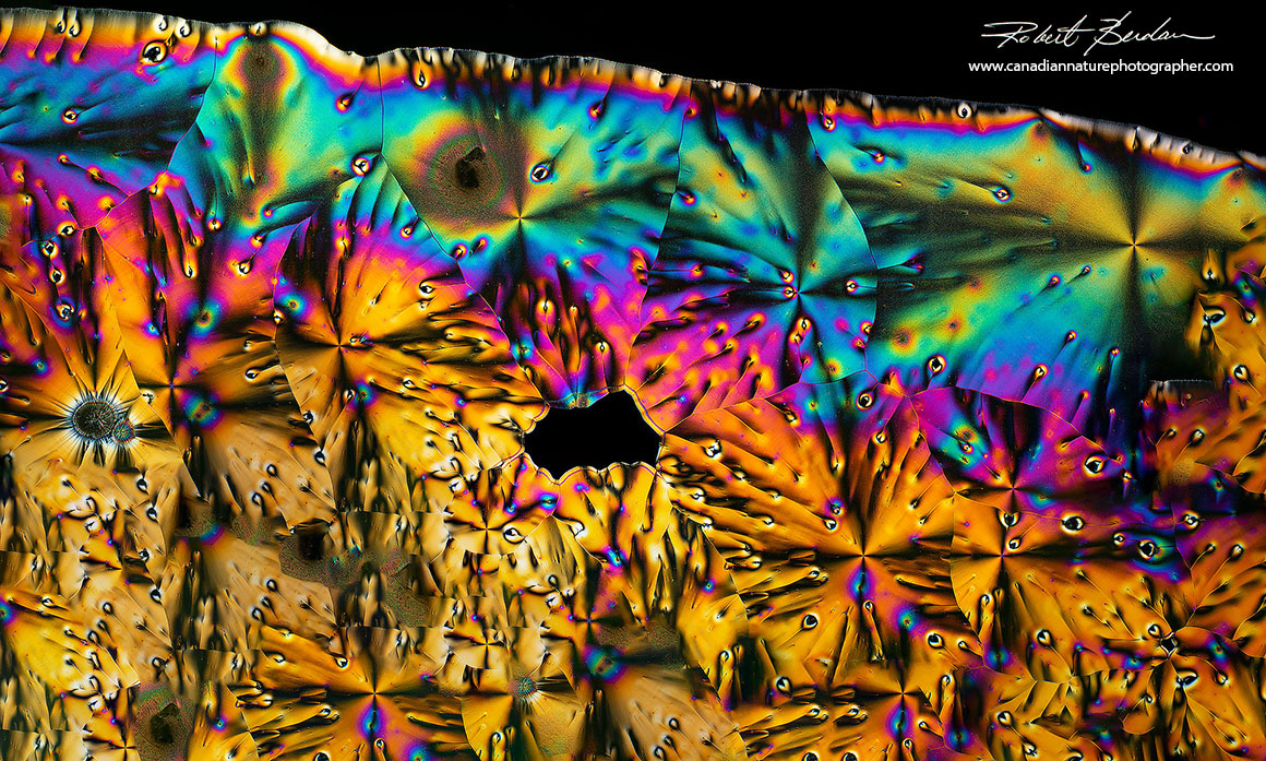
Vitamin C crystal panorama created by stitching 24 images where each individual image was taken at 40X with a polarizing microscope. Stitching images permits the ability to create really large prints and capture wide fields of view.
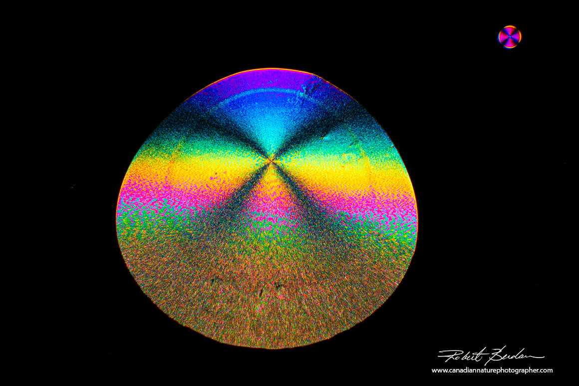
Single crystals of Vitamin C. The crystal starts to grow from the center of the cross. The dark cross represents the regions where polarized light is blocked and is called a Maltese cross - hence it matches the black background (L.M. Volker-Pop 2014 - PDF). Note Ascorbic acid is the form of vitamin C found naturally in food. It has good bioavailability but some people find it too acidic on their gut and can't tolerate high doses. Bioflavonoids are beneficial plant compounds often added to vitamin C supplements (R.Bartholomew nutriadvanced.co.uk).
Vitamin C crystal showing one side smooth while the other side appears plant-like. 40X polarized light microscopy.

Image above of Vitamin C crystals that form bracket fungus-like rings (see picture of Turkey tail polypore). You can see that variations during crystallization can result in very different looking crystals even from the same starting solution. 40X polarized light microscopy.

Vitamin C crystals by polarized light microscopy exhibiting different colours, textures and shapes. 100X Polarized light microscopy.
Menthol Crystals
I used menthol as an anesthetic in my research laboratory when I studied brain cells in snails. Snail brain cells are very large and have remarkable regenerative abilities. The menthol was dripped into the pond water to anesthetize the snails before I dissected them and then extracted individual nerve cells which I grew in cell culture - see scanning electron micrographs of their nerve cells.

Menthol crystals formed by the melt method and viewed by Polarizing microscopy 40X.

Menthol crystals by melt method. Polarizing microscopy 40X

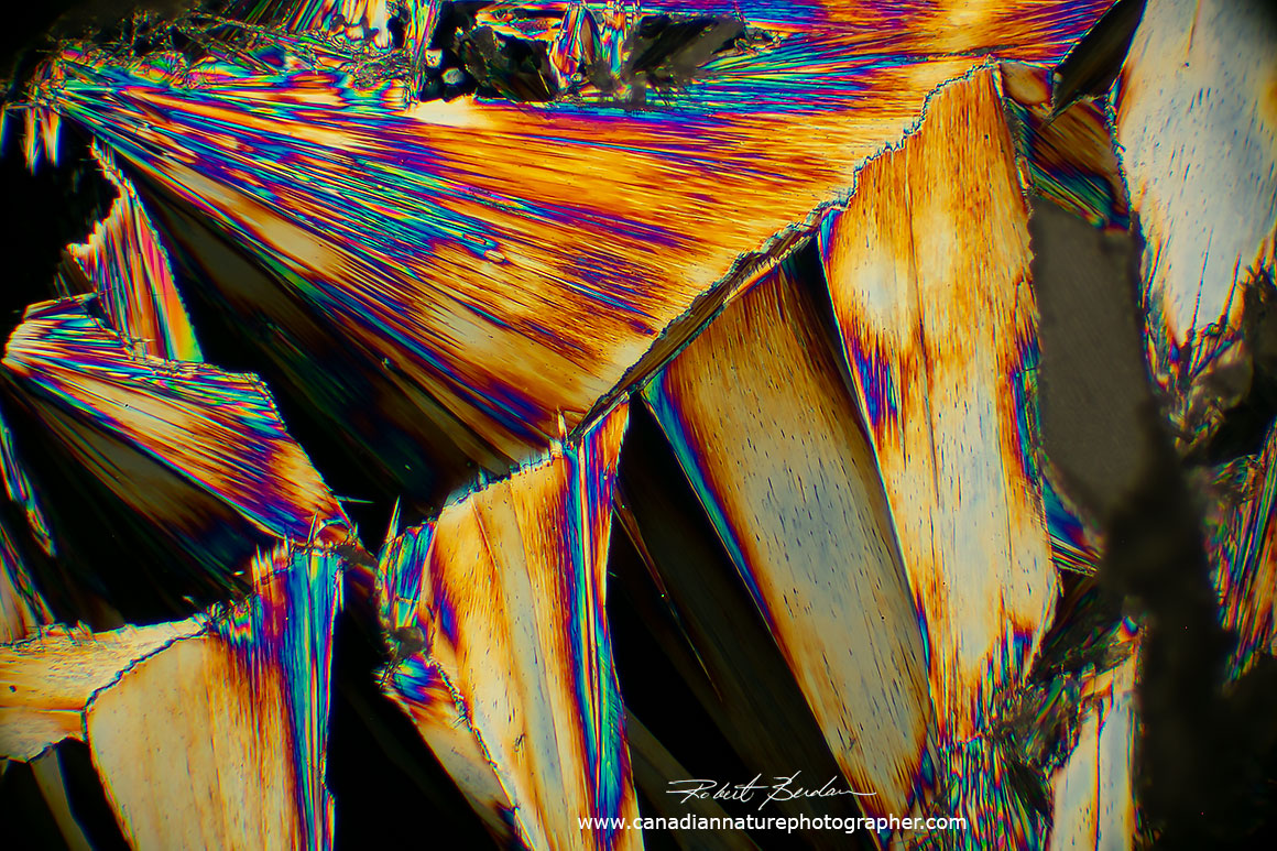
Above image shows that as a crystal increases in thickness we begin to see colours produced by interference which I call the "rainbow effect". The grey areas of the crystal are the thinnest regions and only provide grey to white interference colour.As the thickness of the crystal increase the colours become less saturated - called higher order interference colours. Polarized light microscopy 40X .
Caffeine Crystals
Caffeine is found in coffee, tea, colas and many other food products and is the most widely used psychoactive substance in the world. Caffeine acts as a stimulant which is one reason so many of us like to ingest it. From an athletic point of view it can increase our reaction time by about 3% and our endurance by up to 20%. Too much caffeine though can cause anxiety and reduce athletic performance. Exactly how much one needs to ingest depends on how much is consumed on a daily basis and idividual's tolerance. Some individual's hands exhibit tremors after drinking too much coffee. Tremors occur because caffeine increases the permeability of the sarcoplasmic reticulum in the muscles and makes them more permeable to calcium. Calcium leaking from this reticulum to the muscle causes muscle tremors.
Our endurance is also affected by caffeine by sparing the use of muscle glycogen (e.g. W.J Pasman et al. 1995). For more details see the literature (e.g. Caffeine Use in Sports 2008 - PDF). Caffeine may also increase your heart rate by about 10 beats per minute so its not recommended for sports requiring accuracy like shooting or individuals that may be anxious in competition. The affect of caffeine varies with individuals and in small amounts can improve performance. Never take pure caffeine it is toxic). I used to drink ice tea during competition when playing racquet sports when I was younger.
Caffeine exhibits anhydrous polymorphism, the ability of a solid to exist in more than one form of crystal (R. Carlton 2011). I use two methods to make caffeine crystals 1) dry the solution of pure caffeine in water onto a slide and they will form pointed needles. 2) The second method is to place some caffeine on a slide with a coverslip and heat it gently until it melts (227 °C) then allow it cool for a few minutes. Melted caffeine will often form crystal-sheets resembling feathers with embedded hexagonal patterns resembling snow flakes (see below).
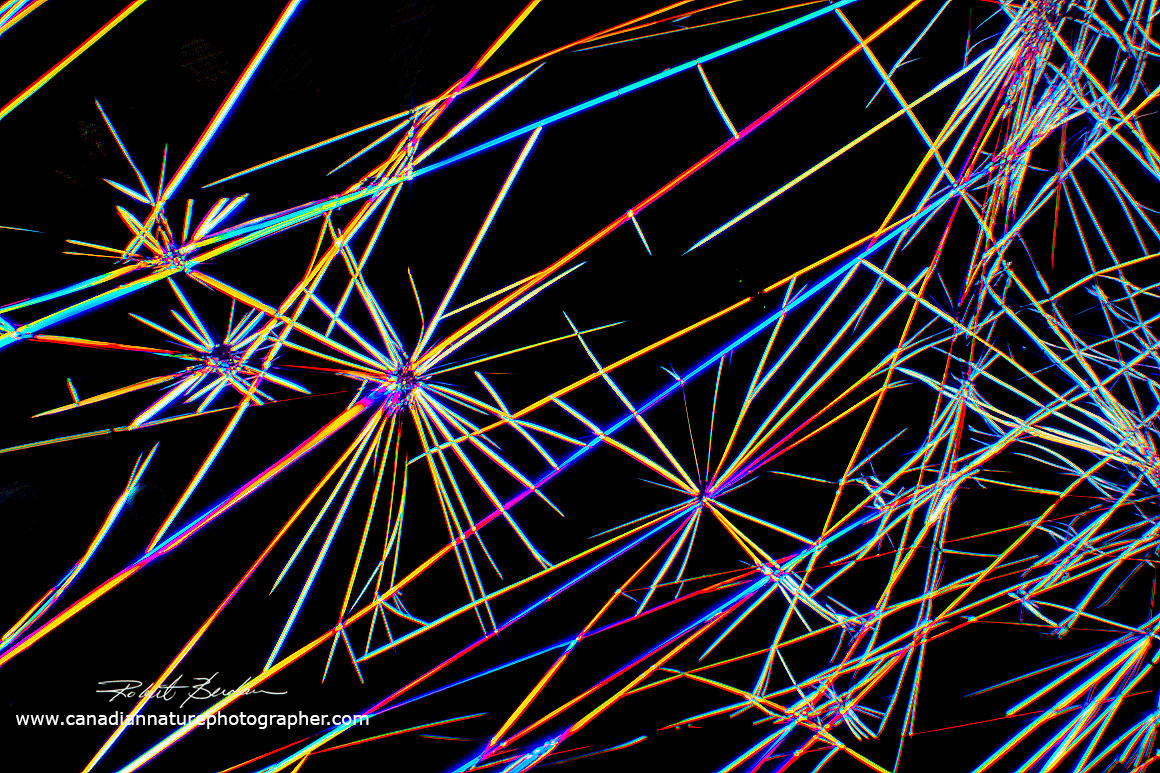
Caffeine crystals often form needle-like crystals when a saturated solution in water is dried onto a microscope slide. 100X polarized light microscopy.
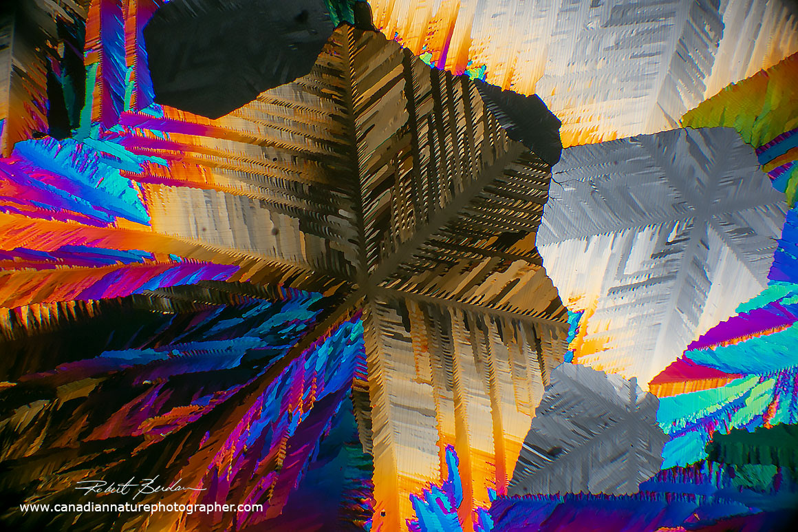
Note the snowflake-like hexagonal patterns in these caffeine crystals using the melt method. 40X polarized light microscopy.
Unusual caffeine crystals that are leaf-like produced by the melt method. Polarized light microscopy 40X.

Above note the hexagonal snowflake-like pattern on the lower left corner of these caffeine crystals. These crystals were produced using the melt method 40X Polarized light microscopy.
Caffeine crystal-sheets are common when melting caffeine on a microscope slide under a coverslip. Overlaying the crystals with a coverslip helps restrict the crystals to thin sheets. When heating the slides be sure to wear protective eye-glasses as the coverslip sometimes explodes or blows off the slide during the heating process. Slides prepared using melts or solutions can be preserved by simply storing them in a slide box to protect them from dust. Sometimes you can melt the same sample several times.
I love to make crystals for photomicrography during the winter as collecting pond organisms is difficult due to the ponds being frozen. Photographing crystals with a microscope also makes for an exciting hobby for anyone that has mobility issues or is restricted to home (e.g. during the COVID pandemic). The substances that can be tested using a polarizing microscope are endless and include crystals inside plants (e.g. potato starch grains), wood, hair, minerals etc. A wide variety of chemicals can be purchased online, but always read about the chemical before you start experimenting with them so you are aware of any dangers they might pose e.g. some can be explosive, or they may form toxic fumes (e.g. Benzoic acid). Children heating chemicals should be supervised by an adult. Vitamin C is good chemical for children and anyone to start experimenting with and does not require any heating. Anyone interested in learning more about microscopy can visit me in Calgary - I am also available for training at an hourly rate to teach adults microscopy and photomicrography in my home laboratory. I am happy to provide 1-2 hours of free instruction on microscopy to students (ages 12-17) with under the supervision of a parent or adult - once the pandemic is over!

My home microscopy lab showing some of my microscopes used for taking pictures. The Motic BA310 polarizing microscope used to take pictures for this article is 2nd from the right. All my microscopes are attached to the computer for recording photographs and movies. You can also hire me to photograph or make movies of anything with a microscope including commercial products, food, drinks etc - see my other web site Science and Art for some additional examples of filters, tiles, wood etc by both light and scanning electron microscopy.
Summary
Crystals are relatively easy to produce from many substances and can form beautiful abstract images. A polarizing microscope is widely used to analyze pharmaceutical drugs, chemicals and in forensic science. By experimenting with safe chemicals like Vitamin C anyone at almost any age can begin to explore the beauty of crystals. Photomicrographs can be taken using a cell phone when starting out. I have been working with polarized light in microscopy for decades and still find it fascinating. The images produced by amino acids are some of my favourite. If you are considering the purchase of a microscope see my article: Tips for Buying a Light Microscope. I occassionally have refurbished microscopes for sale. A new microscope can be purchased starting around $100-$500 and for a few more dollars you can add polarizing filters. If you have questions about microscopes or viewing specimens feel to email or call me with your questions. RB
Crystals as Art. All my photographs can be purchased as digital images for printing and personal use starting at only $20 - see my pricing. I can provide images 24 x 36 inches and larger.
Note: Educators and students may use my web images freely for reports and teaching, all other uses or for commercial use please contact me. If you use my images I appreciate attribution and a link back to this web site. Note magnifications are approximate on digital devices - 100X means taken with 10X objective, 200X with a 20X objective, 400X with a 40X objective. Disclaminer: I do not receive money from Amazon for mentioning their products. The products, microscopes, cameras and chemicals are those that I have purchased and they work well for me.
References & Links
Do it yourself Polarizing Microscope - Microbehunter forum
Do it yourself Polarizing Microscope using Vitamin C - Youtube video
Crystal Growth - Under the microscope - Youtube video
Y. Joshi - Beautiful Chemistry: Amino acids under Polarized light - Youtube
Vitamin C crystals by Polarized Light Microscopy - R. Berdan - Youtube video
Polarization microscopy - Youtube video
Beautiful Chemistry - Glycine crystals growth under a polarized light microscope - Youtube video
Abstract Fluid art created using paint that resembles crystals - Youtube video
Amazing acrylic pour Flower painting - resembles flower crystals in polarized light - Youtube
Fluid Art Bloom with with Amazing colours - Youtube
Earth Optics Video 2 Cross polarized Light Microscopy (watch entire series) - Youtube video
Conoscopic Figures - Youtube video
Websites, Papers and Books
C. Autotte (2020) Playing with DIY Wave Plates - PDF
R. Berdan (2018) Photomicrography a Marriage of Science and Art microscopy-uk.org.uk - PDF
J. G. Delly (2017) Essentials of Polarized Light Microscopy and Ancillary Techniques - available on Amazon and from McCrone - Expensive, but it is the best book on Polarized microscopy I have encountered with excellent illustrations and photographs - it is written for teaching.
A.F. Frandsen (2016) Polarized Light Microscopy - NASA - PDF
Lordana M. Völker-Pop (2014) Optical methods in Rheology: Polarized light imaging. Chem.Listy 108:697-724 - PDF.
D. D'Orazo (2014) Under the microscope, your drink is a colorful piece of abstract art - photos by Michael W. Davidson using polarized light microscopy.
R.A. Carlton and R. Allen (2011) Pharmaceutical Microscopy. Caffeine crystal properties - p 228-235. Springer NY.
A.Shalmashi and F.Golmohammad (2010) Solubility of Caffeine in water, ethyl acetate, ethanol, carbon tetrachloride, methanol, choloroform, dichloromethane, and acetone between 208 and 323K. Latin America Applied Research 40: 2 83-285 - PDF
Brian Johnston (2005) A Demonstration of the Use of Quarter & Whole wave Plates to alter the colouration of Polarized light photomicrographs - www.microscopy-uk.org.uk
E. Wood (1974) Crystals and Light: An Introduction to Optical Crystallography 2nd ed. Dover Publications, N.Y. - available on Amazon.
R. Weaver (2003) Rediscovering Polarized Light Microscopy. American Laboratory pg 55-61 - PDF
Shiroshita et al. (1992) Method for crystallization of amino acides - Patent
A. Watanabe et. al. (1985) Study of Crystalline Drugs by Means of a Polarizing Microscope . VI. Polarizing Microscopy of Drugs acting on the Nervous System and on Individual Organs listed in the Japanese Pharmacopoeia X. Chem. Pharm. Bull. 33: 1599-1608 - PDF
Digicam control software to capture images from a DSLR onto your computer (free).
Home made wave plates - Microbe Hunter
Understanding Wave plates and Retarders - Edmund Optics
Motic BA310 POL microscope - website with microscope specifications and pricing
R. Berdan The Art & Science of Photomicrography with Polarized light
Jearl Walker (1977) The Amateur Scientist - Scientific American. Studying polarized light with quarter wave and half-wave plates of one's own making. This article started me using wave plates years ago.
T.J. Fagan and M.D. Lidsky (1974) Compensated Polarized Light Microscopy Using Cellophone Adhesive Tape. Arthritius and Rheumatism 17: 256-262 - PDF
K. Hayashi et. al (1966) Solubilities Studies of Basic Amino Acids. Agr. Biol. Chem. Vol 30. p 378-384 - PDF
Isolation and Sublimation of Caffeine from Tea Leaves - experimental procedure - PDF
Ms. R.R. Shinde and Prof N.H. Shinde (2017) Extraction of Caffeine from Coffee and preparation of Anacin drug. Int. J. Eng. Res. and Techn.10:236-239 - PDF
M.M.Raith et. al (2012) Guide to Thin Section Microscopy 2nd Edition - about polarized light microscopy of mineral sectins - PDF
Motic Microsocopes - located in Vancouver, BC sells microscopes - moticmicroscopes.com
Quality Microscopes located in Calgary & sells Zeiss Stereomicroscopes - qualitymicroscopes.ca
Authors Biography & Contact Information
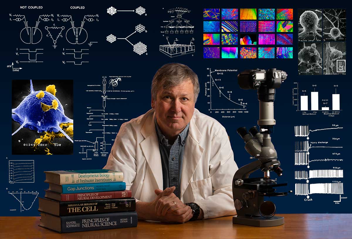
Bio: Robert Berdan is a professional nature photographer living in Calgary, AB specializing in nature, wildlife and science photography. Robert retired from Cell\Neurobiology research to pursue photography full time many years ago. Robert offers photo guiding and private instruction in all aspects of nature photography, Adobe Photoshop training, photomicrography and macro-photography. Portrait of Robert by Dr. Sharif Galal showing some examples of Robert's science research in the background.
Email at: rberdan@scienceandart.org
Web sites: www.canadiannaturephotographer.com
www.scienceandart.org
Phone: MST 9 am -7 pm (403) 247-2457.