
Beyond the Naked Eye - Microscopy & Photomicrography
Where Science Shapes Art
by Dr. Robert C. Berdan, June 10, 2026
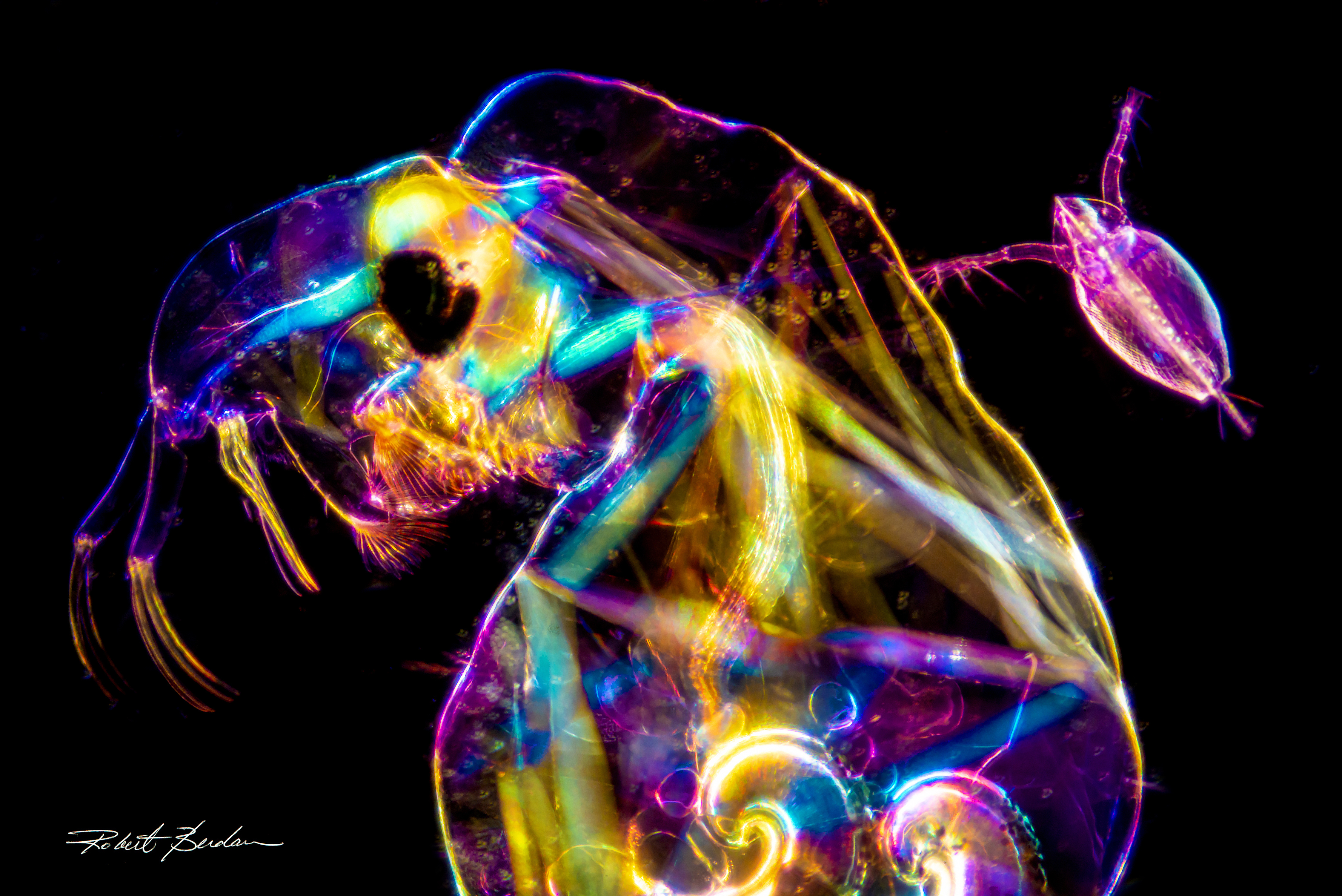
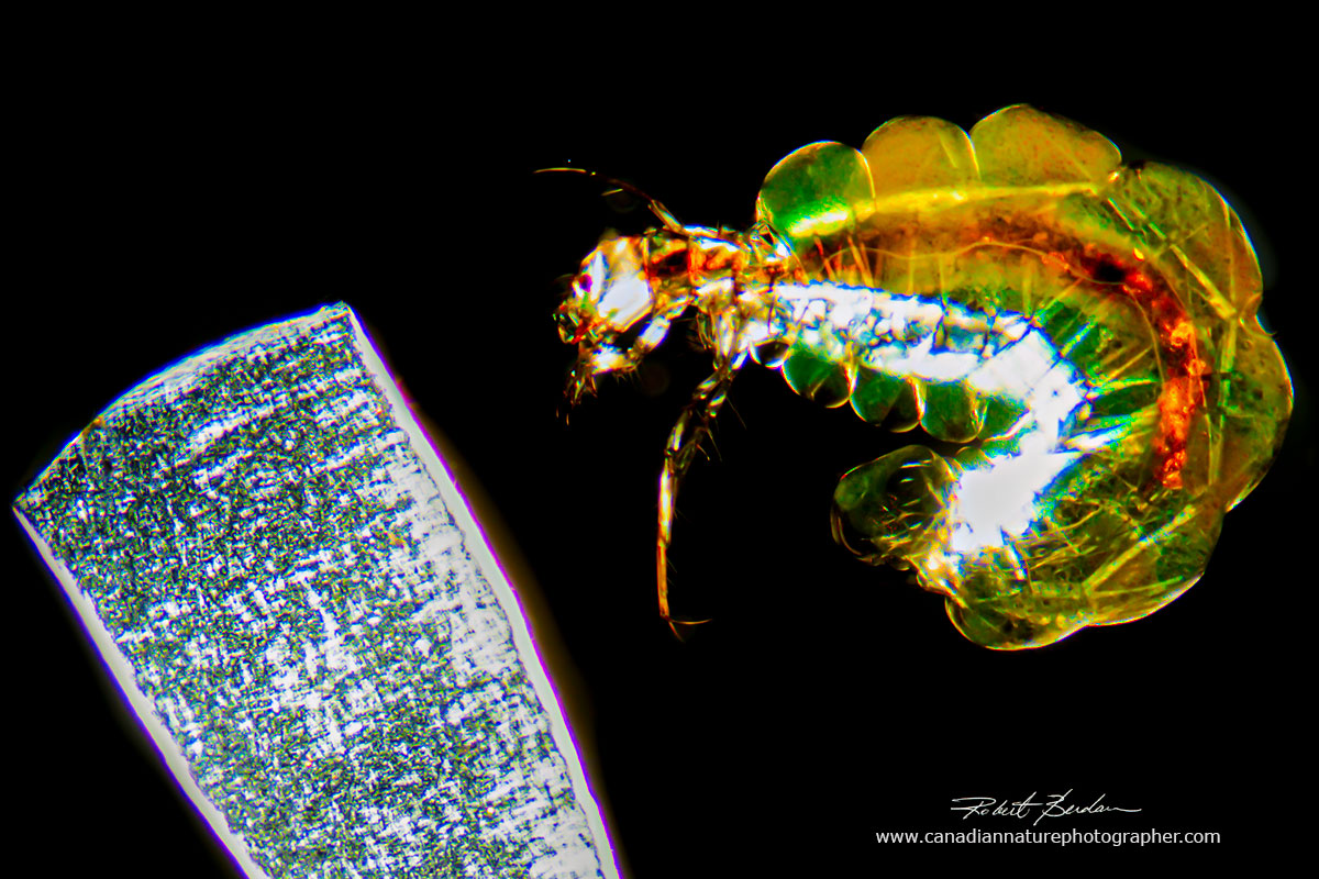
Chaoborus, a midge fly larva is shown alongside a water flea (Daphnia) in the upper right, viewed at 100X magnification shown by a combination of polarizing and dark-field light microscopy. Both organisms are just a few millimetres in size and common in freshwater.
Introduction
Microscopy has never been more accessible than it is today. What once required specialized laboratories and expensive equipment can now be explored with tools available to almost anyone. A simple smartphone, paired with even a modest light microscope, allows students, hobbyists, and even children to record and share subjects from the microscopic universe. The barrier to entry has never been lower.
In this article, I present a selection of my photomicrographs captured over the past fifty years. My passion for microscopy and photomicrography has remained undiminished throughout this time, driven by the remarkable ability of the microscope to reveal structures, patterns, and organisms otherwise invisible to the human eye. As one of the most important instruments in the history of science, the light microscope continues to evolve, and is now capable of visualizing features at the molecular scale.
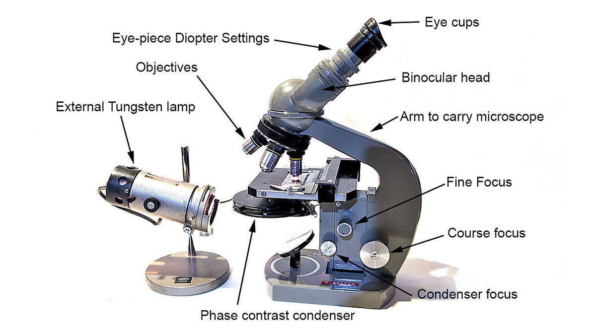
Above is a picture of my light microscope from the 1970s (Olympus). By attaching a camera it is capable of capturing images from 10 to 1000X. and I still use it today.
A used microscope like that shown above can be purchased for only a few hundred dollars, and high quality images or videos can be recorded simply by aligning a smartphone with the eyepiece. This accessibility has opened the door for anyone to explore the micro-universe, investigate scientific phenomena, and even create compelling visual art. With minimal equipment and a sense of curiosity, the microscopic world has become available to all.
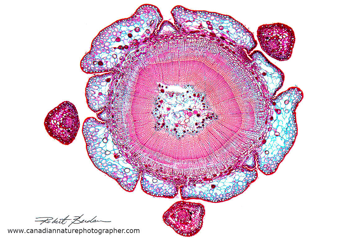
Taxus canadensis stem cross section, commonly known as the Canada yew is a low-growing, needled evergreen shrub native to central and eastern North America. Image was photographed from a prepared microscope slide which was stained. 25X bright field microscopy.
Images courtesy of Wikipedia.
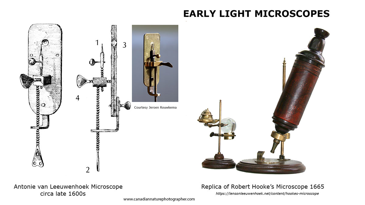
Two key pioneers of early microscopy were Antonie van Leeuwenhoek, who first observed microorganisms in pond water, and Robert Hooke, who discovered plant cells. Their discoveries helped establish that all living organisms are made of cells. Van Leeuwenhoek, who built his own single lens microscopes, is known as the Father of Microbiology. Their work has been well documented by microscopist and science historian Brian J. Ford. See the “The Leeuwenhoek Legacy” in the reference section.
In 1970, the practical limit of resolution for an optical light microscope was approximately 0.2 micrometres or 200 nanometres. Some of the newest light microscopes today are called super resolution microscopes. Several scientists—Eric Betzig, Stefan W. Hell, and William E. Moerner received Nobel Prizes in 2014 for breakthroughs that overcame the diffraction limit of light, allowing biological structures to be seen at the nanoscale (one billionth of a meter) with a light microscope.
In 1933, German physicists Ernst Ruska and Max Knoll developed the first electron microscope that surpassed the resolution of the light microscope. Electron microscopes use magnetic lenses to focus electron beams in a vacuum. The development of a scanning electron microscope started around 1942 with Charles Oatley and his Cambridge students leading to the first Stereo electron microscope in 1965 for surface imaging. The specimens examined from a scanning electron microscope (SEM) appear in three dimensions and black and white. V.K. Zworykin created a dedicated Scanning Electron Microscope in 1942. SEM microscopes are generally used to study the outside of organisms. Today the images are sometimes colourized using software such as Adobe Photoshop. SEM images are not used to study live material, but they can analyze internal components, such as other elements. Most specimens prepared for electron microscopy must be preserved, stabilized, or coated, a process that generally renders the specimen non-viable.
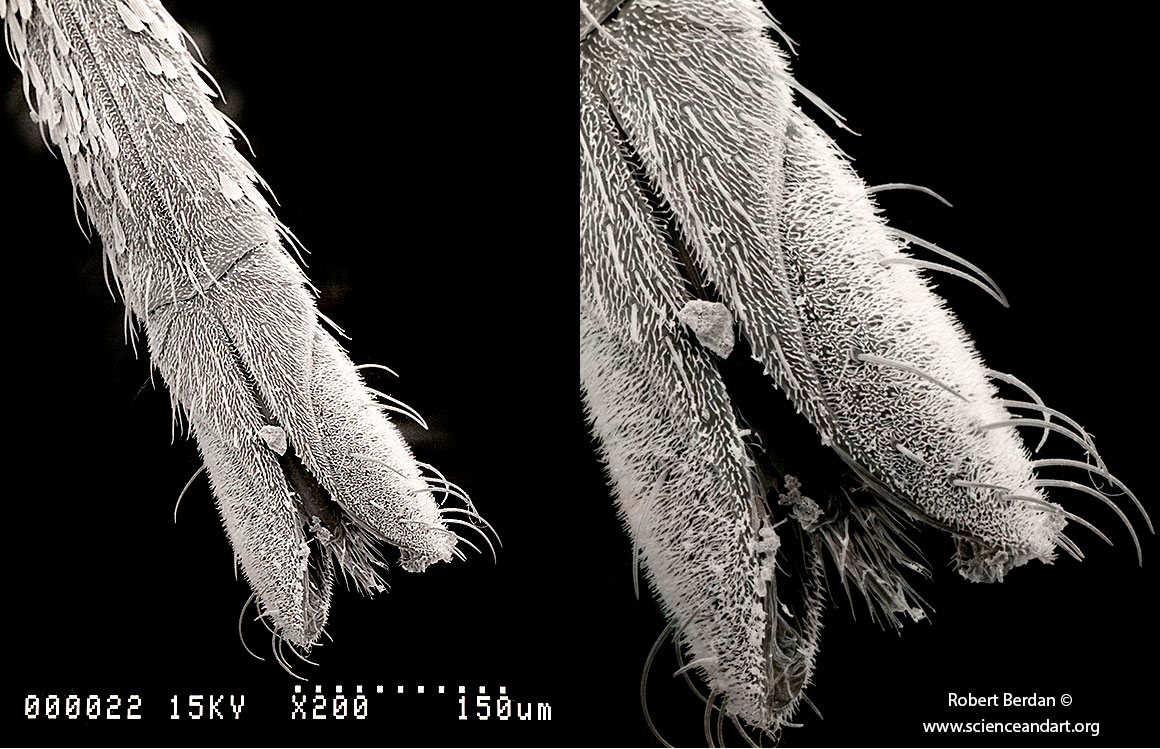
Above are scanning electron micrographs of a mosquito proboscis taken when I was an undergraduate in university. The proboscis is what female mosquitoes use to pierce human skin and draw blood which can result in the transmission of disease. Magnified 200X (left) and 400X (right).
Later in my research, I isolated single neurons and grew them in culture to study how they regenerate and form new synaptic connections. I focused on the central nervous system of freshwater snails because, unlike vertebrate neurons, their neurons are capable of robust regeneration. As part of this work, I isolated individual, identifiable nerve cell bodies and maintained them in vitro. The photomicrographs shown below depict these “identified neurons,” which could be recorded from electrically and would regenerate their axons within days.
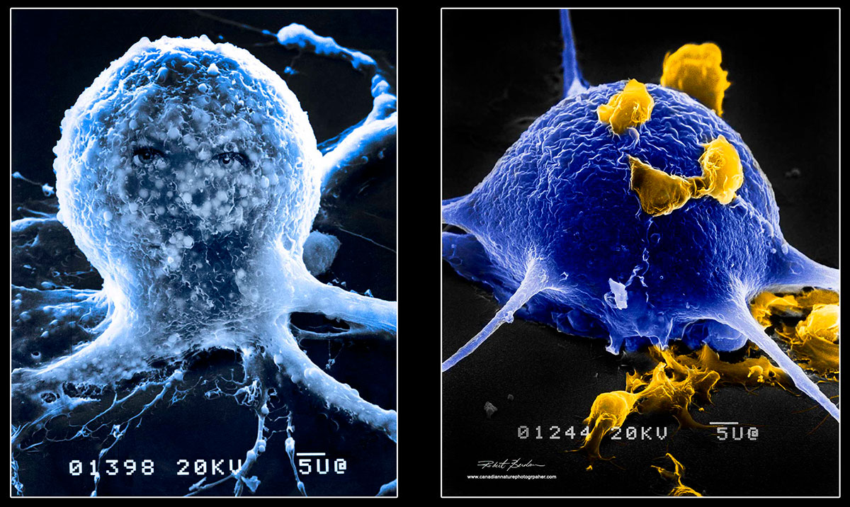
Isolated neurons from a freshwater snail, grown in culture and processed for scanning electron microscopy. The scale bars on the photographs labeled “5u@” indicate a length of 5 microns on the scale bar. One micron equals 0.001 millimeters.
One of the neurons above appears to have “eyes,” though these were added as a playful prank to attract attention during a scientific meeting. In the second image, the yellow structures attached to the neuron are hemocytes (glia equivalents) in snails. Both images were false coloured using Photoshop and magnified approximately 1,500X. These giant neurons measure between 1 and 2 millimeters in diameter.
The goal of my research was to examine how these neurons grow, establish new synapses, and determine what molecular or cellular factors trigger regeneration. Ultimately, I hoped to identify mechanisms that might accelerate or enhance the nerve regenerative process in human cells.
Tardigrades
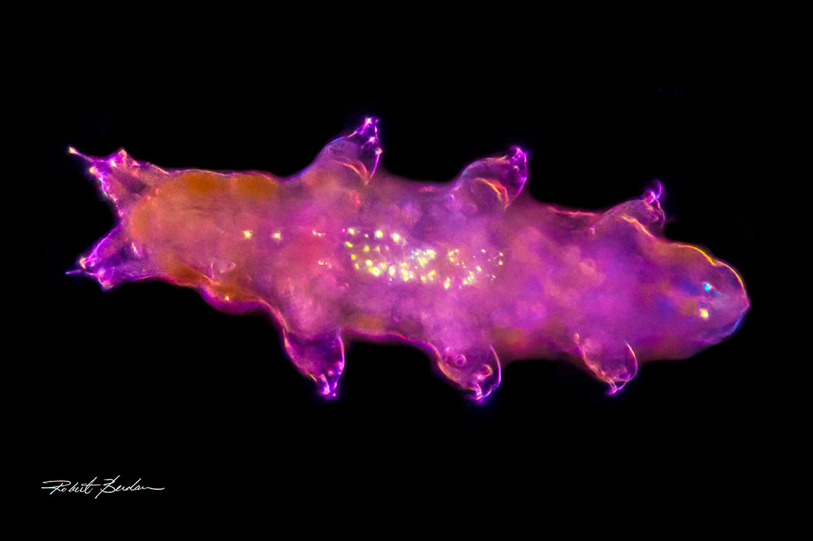
Live Tardigrade found in my backyard and photographed with a polarizing light microscope and dark-field microscopy, 200X. They have 8 legs, 2 eyes, and can survive extremely harsh conditions.
In 2018, I began searching for tardigrades—also called “water bears.” These eight-legged micro-animals are less than a millimeter long and commonly found in lichen, moss, and pond water. Tardigrades are remarkable for their ability to enter a cyst-like state known as a tun, allowing them to survive desiccation, radiation, extreme temperatures, and years of suspended animation in a condition called cryptobiosis. Protein extracts from tardigrade cells have been shown to protect human cells in culture from radiation damage which has attracted interest from NASA, as such molecules may one day help safeguard astronauts during space travel or protect patients undergoing radiation therapy.
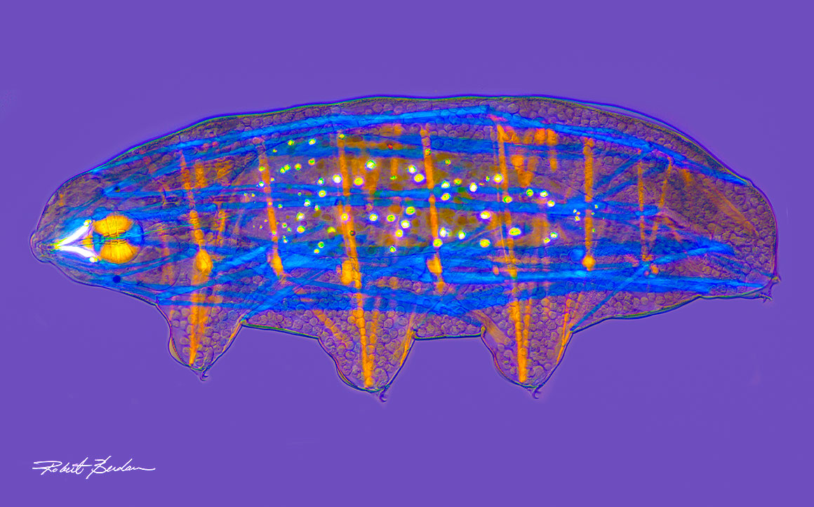
Tardigrade photographed with a polarizing light microscope. The muscles are revealed as blue and orange colours and their stylet which is used for feeding on plants, is located on the left side. This picture is a panorama of 5 separate pictures stitched together and aligned. 200X.
Although tardigrades are nearly ubiquitous in nature, they require a microscope to be seen clearly. Several species live in my backyard, and some individuals I collected carried attached ciliates. When I posted images of these tardigrades on my website in 2018, I learned that this was the first documented observation of attached ciliates on tardigrades in North America. A research student from a university in Poland, Karol Walach, contacted me and asked if I could send specimens so he could determine whether the ciliates represented a new species. In return, he shared scanning electron micrographs of the samples. The taxonomic status of the ciliates are still under investigation.
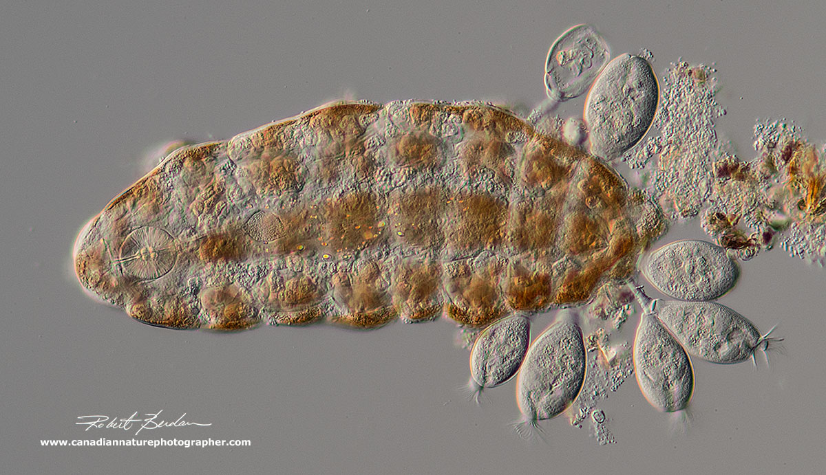
Above is a tardigrade with attached ciliates, photographed at 200X using differential interference contrast (DIC) microscopy. The tardigrade is Ramazzottius oberhaeuseri, and the ciliates clustered near its posterior end appear to be Pyxidium tardigradum. These ciliates feed on bacteria and are not believed to harm the tardigrade directly, though their presence can create drag as the animal moves.
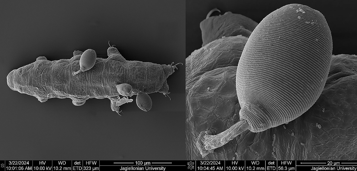
SEM pictures of the Tardigrade with attached ciliates. One enlarged ciliate is shown on the right. These images are courtesy of Karol Wallach, Poland. Each image has a scale bar in microns, 100 & 20 µm.
During my Ph.D. studies in Houston, Texas with Dr. Norton B. Gilula, I was attempting to isolate electrical synapses (gap junctions) from crayfish cells. Below I show a transmission electron micrograph of isolated gap-junction proteins magnified 1 million times (Berdan, 1987). The enhancement was by a technique called “Markham rotation" using dark room printing. Markham first used the technique with electron microscopic images to enhance the rotational symmetry of viruses. The gap junction proteins appear to be made up of six smaller subunits surrounding a 3 nm core or channel (1 nm = 10-9 meters). The image was false coloured and enhanced. Gap junctions allow adjacent cells to exchange small molecules through pores along with electrical current. Gap junctions allow heart cells to "beat synchronously" and are found in many other tissues like smooth muscle, lens cells, liver and some brain cells.
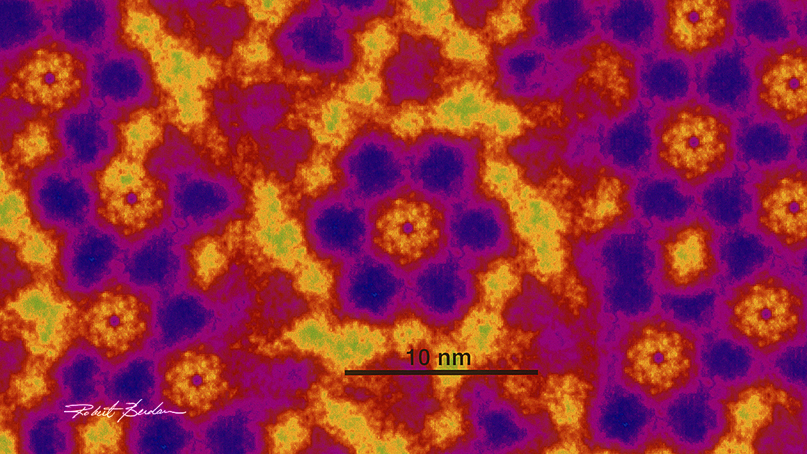
Electron micrograph 1,000,00X. In the center of the photograph there appear to be 6 blue subunits (polypeptides) surrounding a red-yellow channel. These channels allow the exchange of nutrients and electrical current between cells. False colouring shows the protein subunits in the plasma membrane to be seen more clearly. R Berdan 1987.
Electron microscopes are costly and generally only found in universities and research institutions. I have been fortunate to have worked with many types of microscopes in research, but a basic light microscope is something almost anyone can afford.
Light Microscopy
Light microscopes generally come in two main configurations: upright and inverted. Most provide magnification up to about 1000X, achieved by multiplying the magnification of the eyepiece by that of the objective lens. In addition to standard light microscopes, stereo microscopes offer lower magnification, a wider field of view, and a much larger working distance beneath the objectives. They are easier to use, less expensive, and widely employed alongside compound microscopes. Typical stereomicroscopes provide between 1X to 100X magnification and are commonly used to examine insects, fossils, minerals, and used for plant and animal dissections.
Below is a photograph of a butterfly captured with a stereomicroscope. To increase the depth of field, I photographed the butterfly at multiple focal depths and combined the images using a technique known as focus stacking. I also use focus stacking with my compound light microscope to achieve a greater depth of field in high-magnification images.
Most photographers know that depth of field decreases dramatically as you move closer to a subject or there is an increase in magnification. With a camera, this can be compensated for by selecting a smaller aperture—such as f/11 to f/32—to extend the depth of field. However, a light microscope does not offer the same degree of aperture control. To overcome this limitation, multiple images can be captured at different focal planes and merged into a single, sharply focused composite. This process, focus stacking, has become an essential technique for producing high-quality photomicrographs.
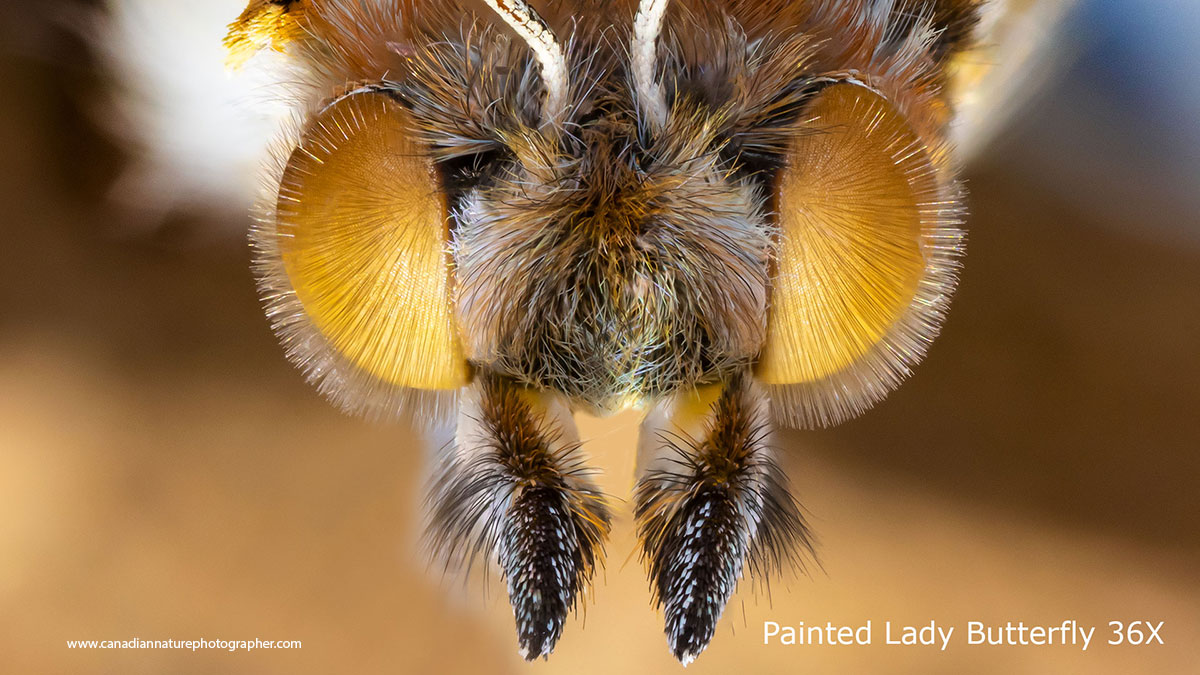
Picture of a Painted Lady butterfly head focus-stacked using a stereomicroscope, magnified 36X.
Most light microscopes provide magnification in the range of 40X to 1000X, although I also use specialized microscope objectives (1X and 2.5X) that offer lower magnifications between 10X and 25X. Achieving magnification beyond 1000X typically requires more advanced imaging approaches, including laser-based systems, fluorescent dyes, digital enhancement, and computer-assisted microscopy. With a standard light microscope, bacteria become visible around 400X or higher. Bacteria are generally small and structurally simple, appearing as spherical, rod-shaped, helical, or spiral forms. Some bacteria species exhibit motility through tail-like flagella, while others contain pigments such as sulfur granules shown below.
Bacterium (Chromatium sp.) found in pond water. This large bacterium was discovered in 1852 by the German naturalist Maximilian Perty. It moves using a single long flagellum, a slender, hair-like structure that propels it through its environment. Magnified 630X by Differential Inference Contrast (DIC) microscopy. Most bacteria are simple in shape like spheres or pill- shaped.
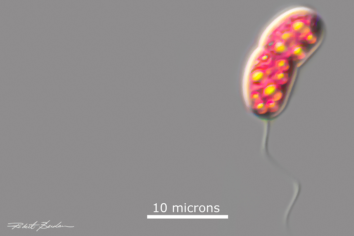
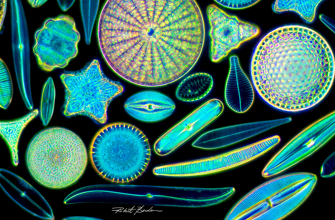
Protists and Diatoms
This photomicrograph of diatoms was captured on 35 mm film back in 1987 with the light microscope shown above. Diatom shells, or frustules, are rigid, porous silica structures that form tiny, ornate glass houses by these single-celled algae. 200X
Some of the most intricate and visually striking microscopic organisms belong to a group known as protists. These are nucleated organisms that don’t fit into the categories of animals, plants, or fungi. Most are microscopic and often single-celled, though some form colonies and others thrive in aquatic environments. Protists make up over a 100,000 species, have a nucleus and are often single celled though some are colonial. They include: diatoms, radiolarians, protozoa, slime molds, euglena, ciliates (e.g. paramecium) etc. Diatoms live in both freshwater and marine environments, and produce oxygen. They have intricately decorated silica shells that form diatomaceous earth after they die, and become a powdery sedimentary rock made from their fossilized remains and are used in silver polishes, tooth-paste, filters and electronic sensors.
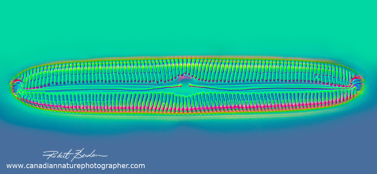
Fresh water diatom shell (non-living), Pinnularia sp. 400X DIC microscopy. Focus stacked.
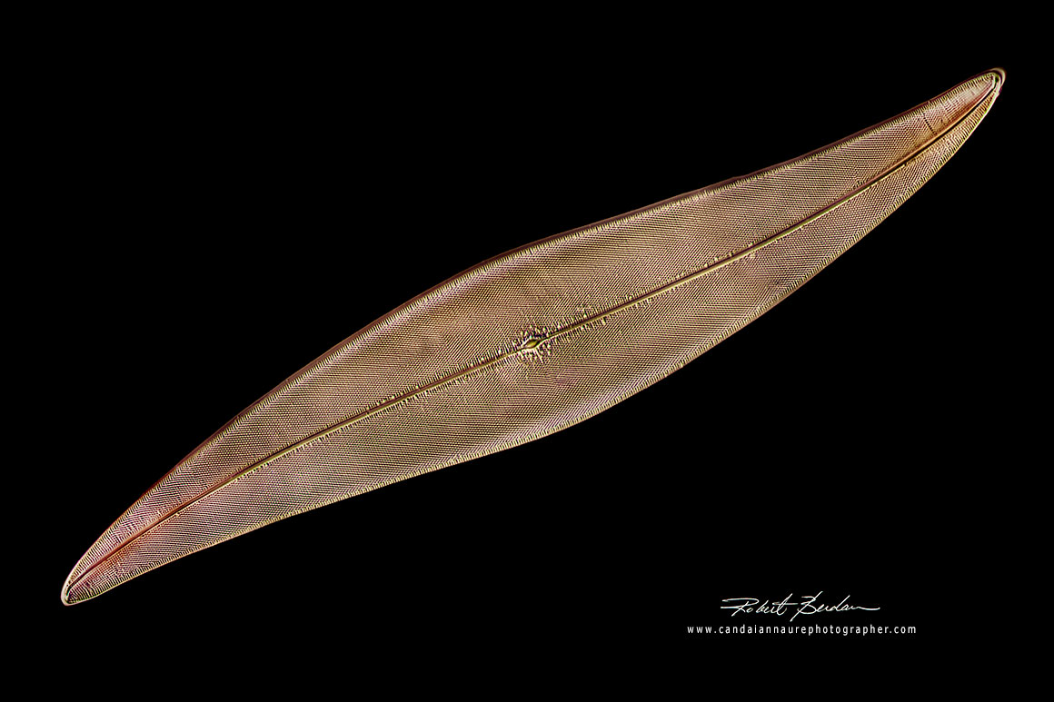
Pleurosigma angulatum is a marine diatom that has long been used to evaluate the resolving power of microscope objectives. It's pores are spaced approximately 0.64 microns apart on average. The specimen shown above was imaged using dark-field microscopy and photographed with a 100X oil-immersion objective.
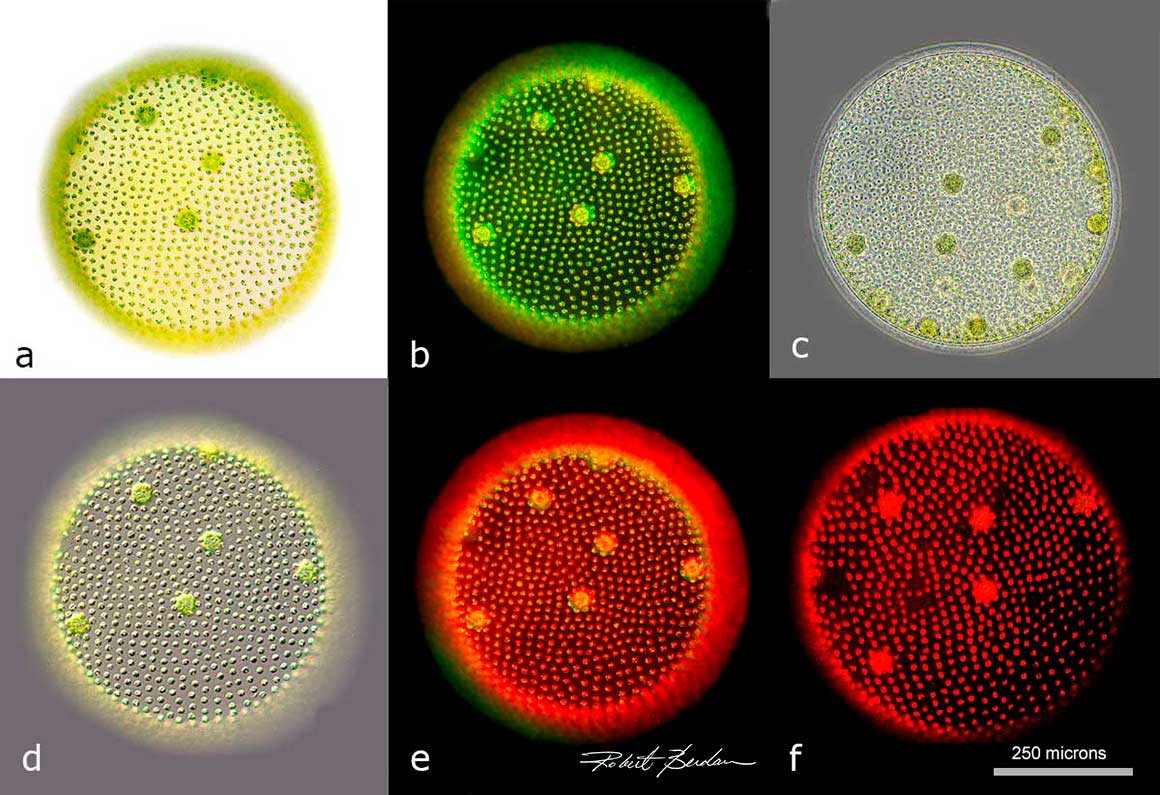
The images above show a colonial alga, Volvox, also a protist and commonly found in pond water. It is capable of forming spheres composed of thousands of cells. The examples illustrate a single Volvox colony viewed by several types of microscope illumination: a). bright-field, b) dark-field, c) phase contrast, d) differential interference contrast, e) Rheinberg lighting, and f) green-light fluorescence. Magnification: 100X.
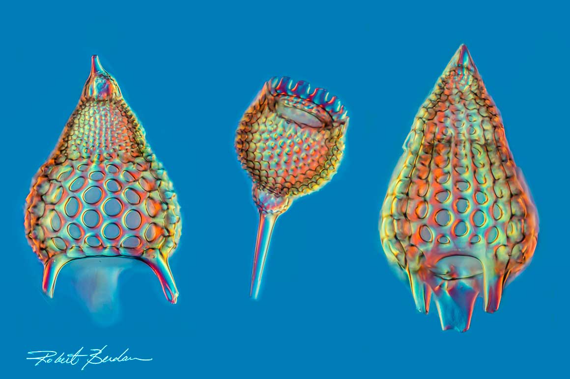
Radiolarian (protists) shells come from marine organisms that build intricate silica structures, much like diatoms. This image shows their detailed architecture at 400X magnification. Image was produced using focus-stacking and viewed with differential interference contrast (DIC) microscopy. Only their their non-living shells are shown.
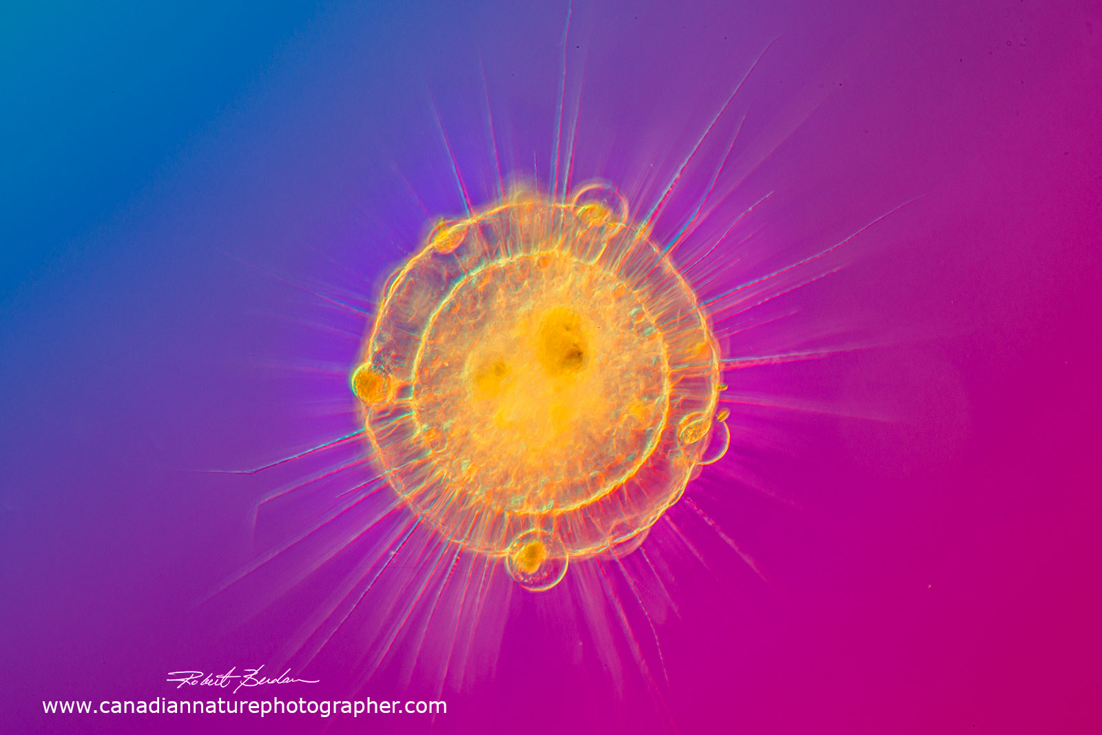 Actinosphaerium is a single celled protist living in pond water and is a genus of heliozoa. It uses spines to impale other protists, and suctions their internal components. 50X DIC microscopy.
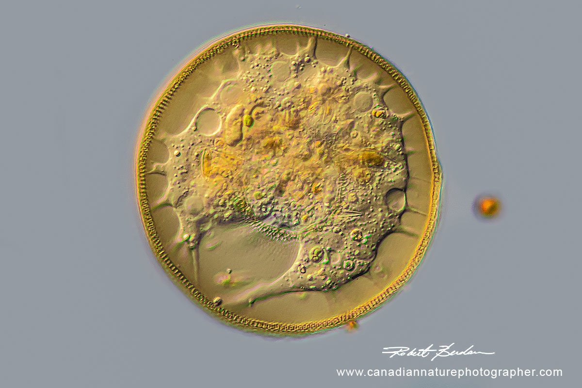 Live testate amoeba. It can feed and move by protruding pseudopodia from its shell and it is found in ponds, wetlands, moss and soil. Differential Interference microscopy (DIC). 400X.
 Single celled ciliates Spirostomum minus (top) and Urocentrum turbo (bottom) - both are common in fresh water ponds (protists). DIC microscopy 100X.
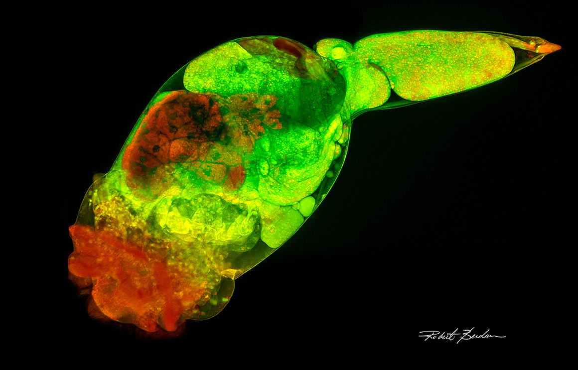
Above Brachionus manjavacas stained with Acridine orange and viewed with a fluorescence microscope. This marine rotifer is widely studied and known for its use as live food in aquaculture and as a model organism in aging studies, toxicology, and endocrinology research. It has a lifespan around 10 days and is made up of approximately 1000 cells. It was alive when photographed and viewed by fluorescence microscopy. 200X.
.
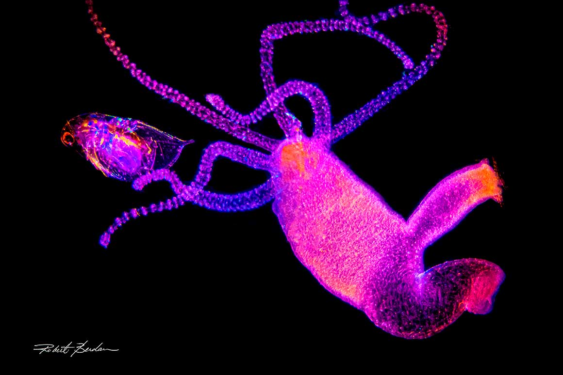 Hydra pond-dwelling organism shown feeding on a Daphnia (left), viewed by polarized light and dark-field microscopy by polarized light microscopy. 50X.
Hydra is a multicellular (approx. 100,000 cells) pond-dwelling organism shown feeding on a Daphnia (left), viewed by polarized light and dark-field microscopy. It reproduces primarily by budding (right side) and is notable for its lack of aging. Hydra captures, paralyzes, and then swallows Daphnia whole. Photograph of live specimens.
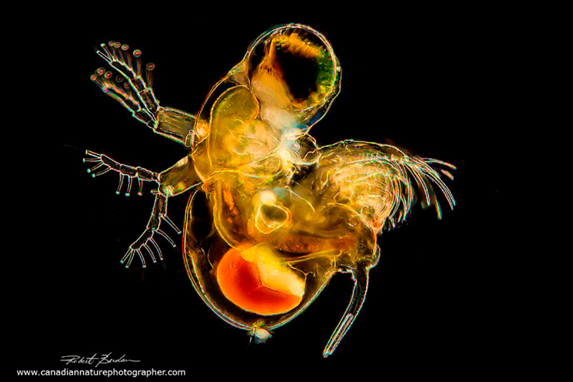
Cladoceran, Polyphemus sp. is also known as a “water flea”and found in fresh water, but unlike other water flea species it is a predator. Dark field microscopy, live specimen 100X.
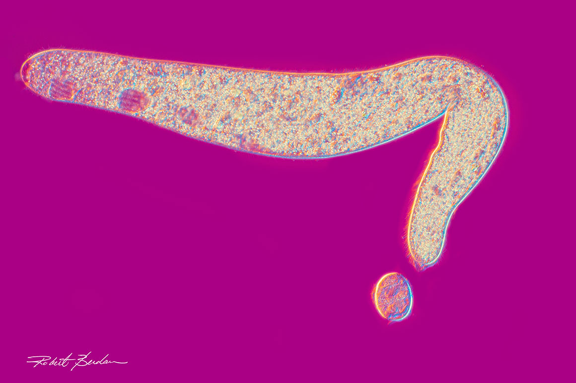
Caddisfly larvae are freshwater insects, and this one belongs to the genus Oxyethira. The white tube on the left is a silk case it constructs and lives in. The larva measures about 2 mm in length. Photographed with dark-field and polarizing light microscopy. 20X
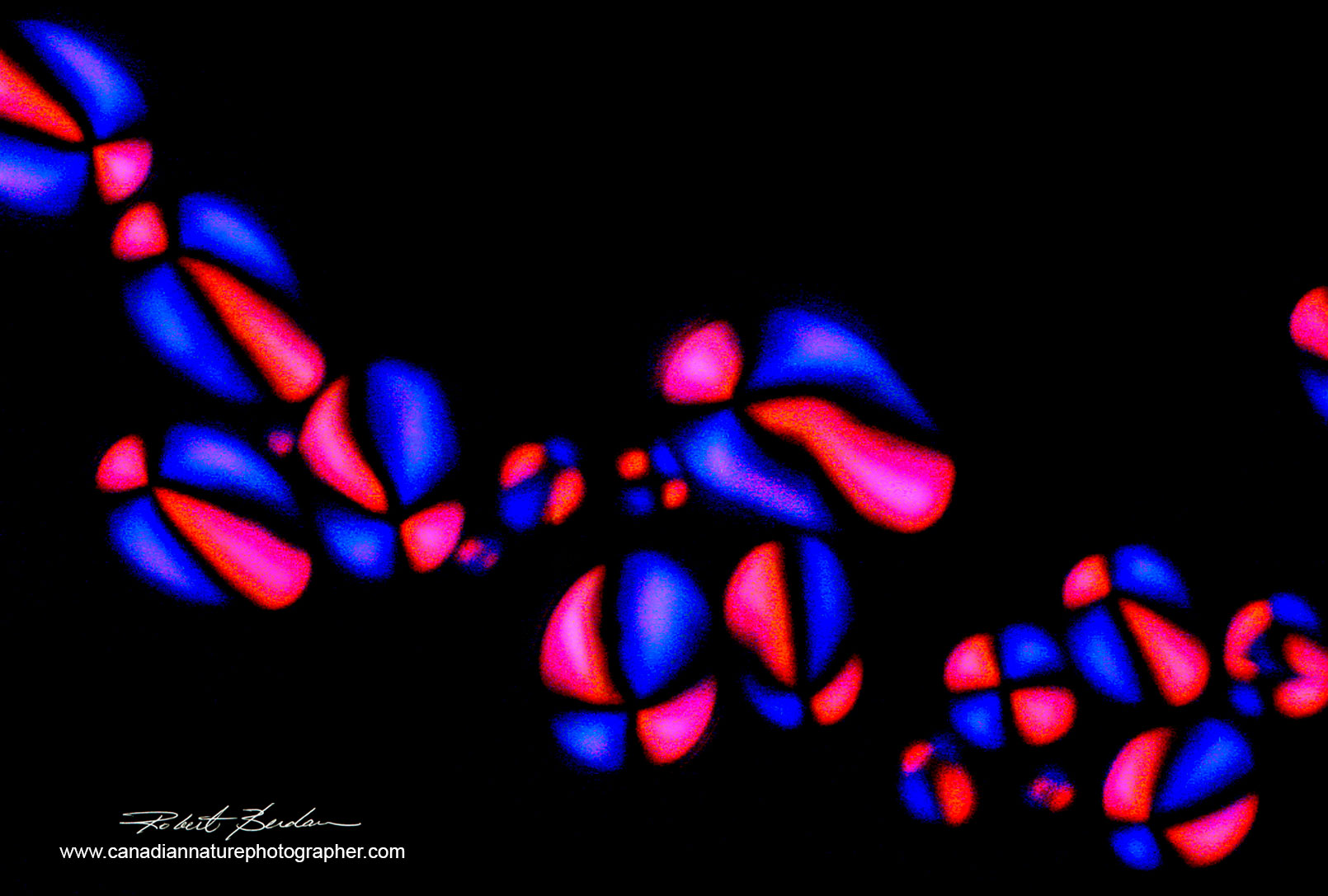
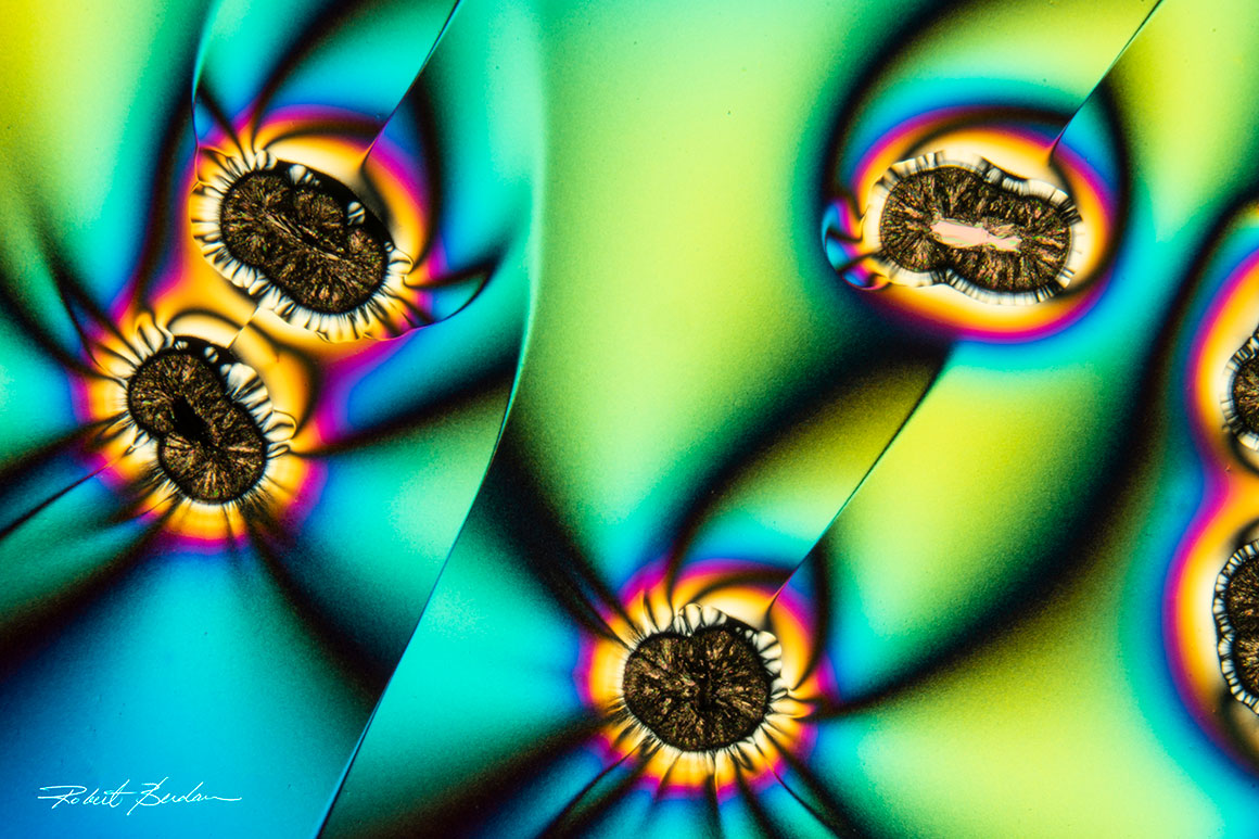
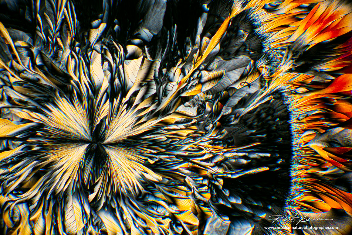
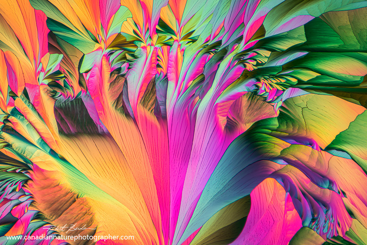
Crystals Viewed by Polarizing Microscopy
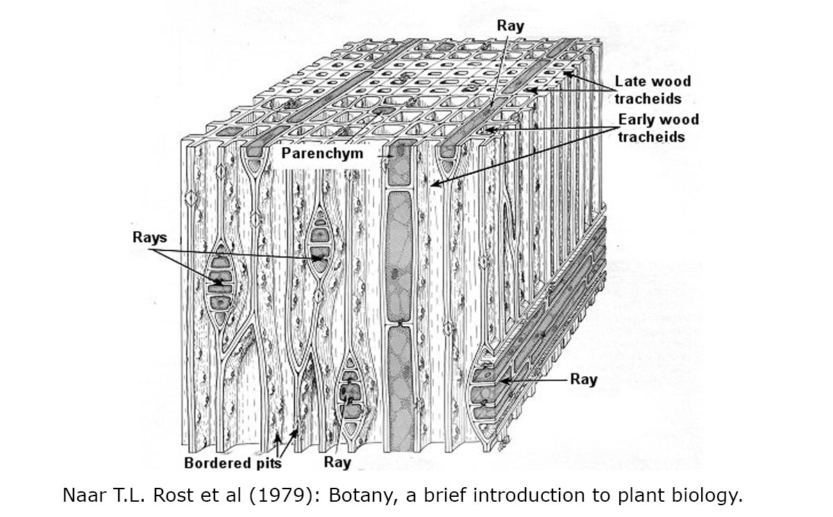
Crystals can display an impressive range of shapes and colors, particularly when viewed with a polarizing microscope. Any standard light microscope can be adapted for polarization by placing a linear polarizer over the light source and a second polarizer, the analyzer, above the objective and over the eyepiece. Additional filters, known as wave plates, can be inserted between the polarizer and analyzer to amplify the colors. Many biological materials (e.g. wood) are naturally birefringent and split polarized light. Polarizing microscopy is used in geology for mineral identification and in forensic science for examining fibers.
 Wood has a semi-crystalline structure. This is a photo of a pine section - xylem tracheids making up wood, photographed with polarized light and a full wave plate.
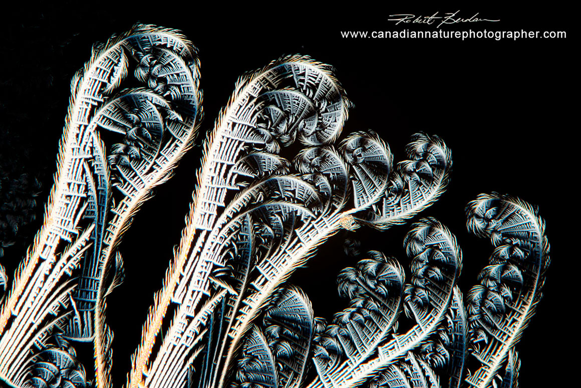
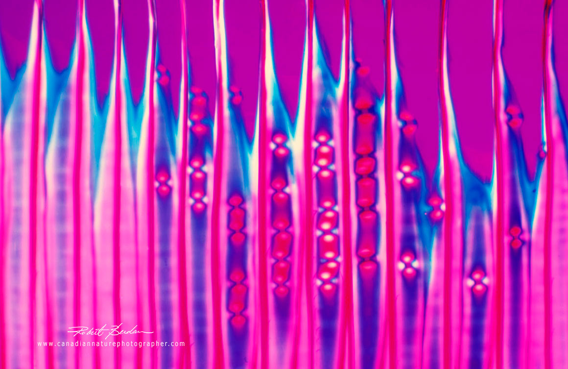
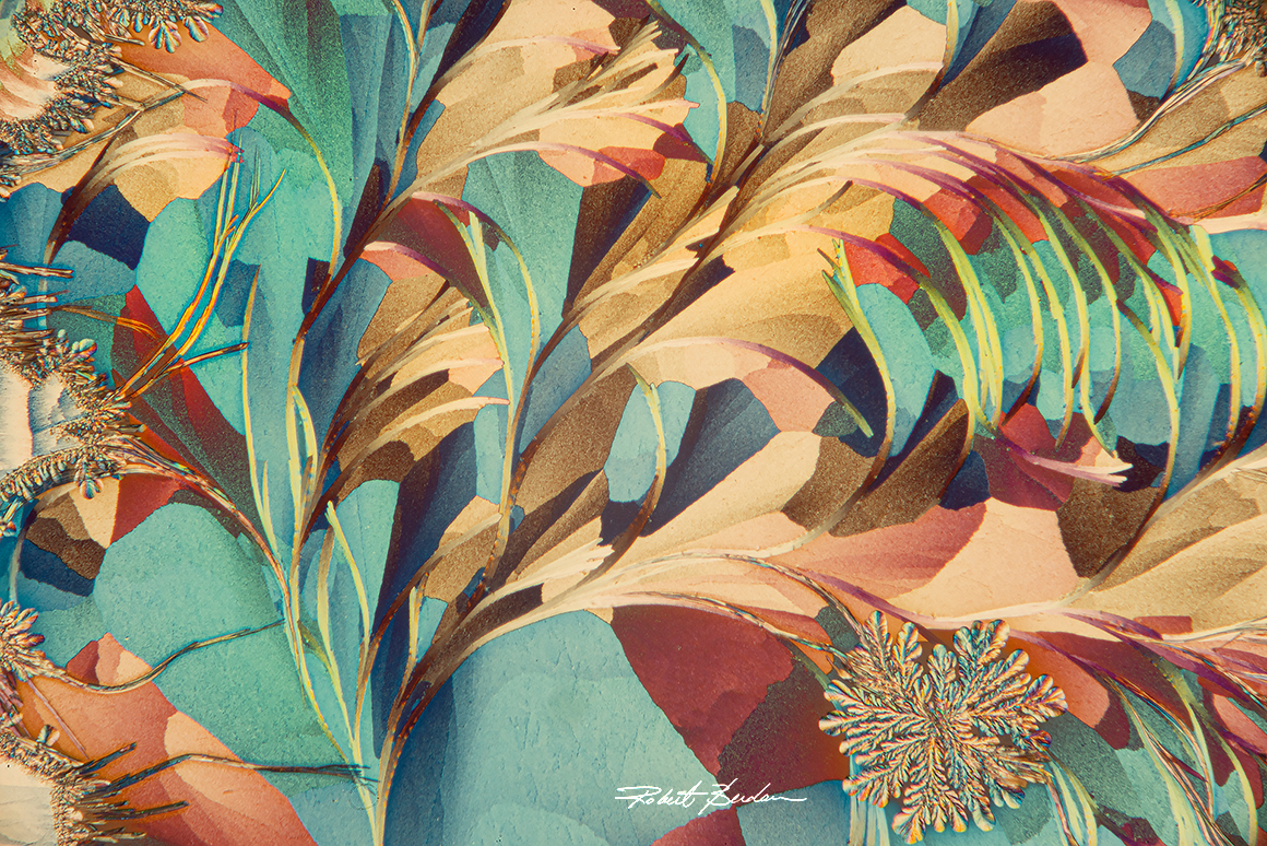
Fern-like crystals from broadacid by polarizing light microscopy. 50X.
Many other substances—such as pharmaceutical drugs, amino acids, and even wine crystals—are birefringent. Most crystals show striking symmetry, and some of the most captivating resemble delicate flowers. Their interference patterns can sometimes be remarkably colourful. Even ice and snowflakes, when viewed under polarized light, can reveal a spectrum of colors created by the way polarized light interacts with its structure.
Potato starch grains examined by polarized light microscopy. Fresh potato pieces were squashed on a glass slide and placed under a cover slip. The starch grains from different species of plants form crystals that have unique shapes. 400X.
Vitamin C crystals shown by polarized light microscopy. 200X
Crystal of amino acids by polarized light microscopy. B–Alanine and L-Glutamine dissolved in ethanol and water and crystallized on a microscope slide by heating. 100X

Wine crystals can develop striking, flower-like patterns under the right conditions. My son and I developed methods for producing crystals from different wines and examined them with polarized light microscopy. 50X.
Crystals of chardonnay wine by polarized light microscopy 50X.
Crystals of ABE callus remover by polarized light microscopy 100X. ABE callus remover, a Polish product, is a chemical blend of salicylic acid, lactic acid, and other chemicals like turpentine.
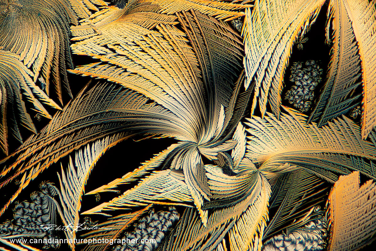 Broad acid crystals in polarized light microscopy 50X. Broadacid is used to remove warts and it is made in Poland. By smearing and diluting it in acetone it offers beautiful art-like crystals.
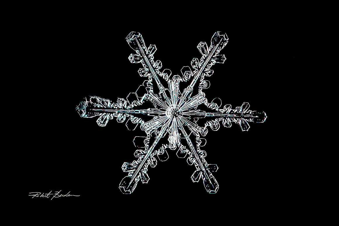
Snowflake - water crystal photographed in in my backyard during winter 25X.
Yeast
Yeast cells are widely used in research, as a single-celled, sugar-eating fungus essential for human civilization. Its main significance is its role in fermentation, where sugars are converted into carbon dioxide and alcohol.
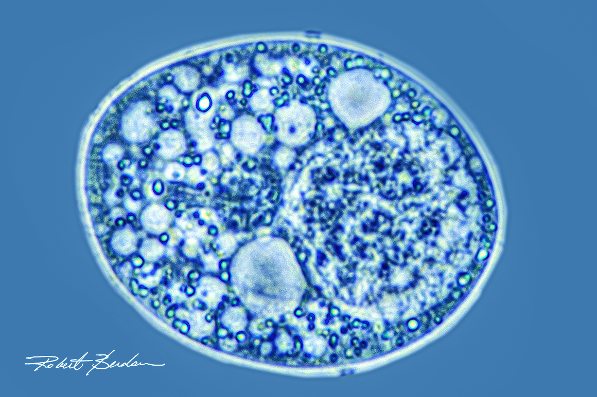
Above is a live yeast cell (Saccharomyces cerevisiae), an organism essential to both baking and brewing. Viewed with phase contrast microscopy, the cell’s nucleus appears on the right side. Image captured at 1000X magnification using a light microscope with an oil-immersion objective. Up until the 1980's this was close to the maximum detail anyone could see in a single living cell.
Today at ~120 nm resolution, scientists can now observe cells with super resolution light microscopes showing the shape and movement of organelles in real time. Careers involving light microscopes are growing, diversifying, and becoming more high-tech, especially as AI, automation, and super-resolution imaging expand what microscopes can do. What might start as a hobby could turn into a career.
Recommendations for Getting Started in Photomicrography
If you’re considering purchasing a microscope, begin by learning as much as you can about the different types and features available. A used microscope is often the best entry point—many modern, Chinese-made models offer excellent optical quality at a relatively low cost. Premium brands such as Zeiss, Nikon, Evident (Olympus) and Leica produce superb instruments, though they are more expensive. For additional guidance on what to look for in a microscope, see my article “Tips for Buying a Light Microscope” on my website.
For photography, I recommend choosing a microscope with a tri-nocular head. Start with a smartphone camera paired with an inexpensive phone adapter; this setup is surprisingly capable and very easy to use. DSLR cameras from manufacturers such as Nikon, Canon and Sony provide higher resolution and greater flexibility, and they can also be used for general photography. Free software for controlling DSLR cameras for a Windows computer is available at Digicamcontrol.com. Apple users should consult their camera manufacturer for compatible capture software. It’s always helpful to seek advice from experienced microscopists, microscopy clubs, or researchers who can offer practical insights. If you have questions about microscopes or photomicrography, feel free to email me rberdan@scienceandart.org —I’m always happy to help others get started in microscopy.
Summary
Microscopes and cameras are hands-on tools that spark curiosity, exploration, and lifelong learning. Because microscopes can be used indoors and in any season, they offer endless opportunities to examine and photograph an astonishing variety of subjects—far more than I can show here. A well-built microscope can last a lifetime, and a wide range of accessories and learning resources are available online. A smartphone is an excellent starting point for photomicrography, and even with simple equipment you can discover new and fascinating microscopic worlds. Enjoy the process of exploring, observing, and capturing the beauty of the micro-universe.
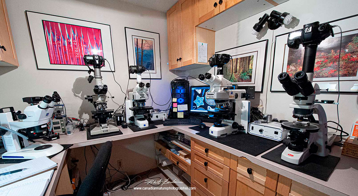
My darkroom was converted into a microscopy room about a decade ago and features both stereo and light microscopes connected to a computer using Digicam control - free software.
Acknowledgements
I would like to thank Marek Mis of Poland for generously sharing his photomicrography on my website several years ago. My thanks also go to Brian J. Ford in England for his inspiration, historical insights, and influential contributions to the field of microscopy. I am grateful to Karol Walach, who provided SEM images of the tardigrades with attached ciliates that I sent him; he recently completed his M.Sc. research on tardigrades. Dr. Bruce Taylor in Canada assisted me in identifying several ciliate species, while Dr. Robert Walsh and Dr. Russell J. Shiel in Australia helped with the identification of cladocerans (water fleas) and rotifers. Finally, my heartfelt thanks go to my wife, Donna, who has supported me in collecting pond samples.
References
R Berdan (1987) Intercellular Communication in Arthropods – Biophysical, Ultrastructural and Biochemical Approaches. From Cell-to-Cell Communication. W. C DeMello. Ed. Plenum and Springer Publishing Chapt. 10. 299-370.
R. Berdan (2017) Focus Stacking, comparing Photoshop, Helicon Focus and Zerene. https://www.canadiannaturephotographer.com/rberdan_focus_stacking.html
R Berdan (2013) Tips for Buying a Light Microscope - Compound, Inverted and Stereoscope and Why you might want to buy a Microscope https://www.canadiannaturephotographer.com/guide_buying_microscope.html
R Berdan and B. Berdan (2020) Cell Phone Cameras, Dedicated Digital Cameras and Digital Single Lens Reflex Cameras for Photomicrography https://www.canadiannaturephotographer.com/cellphones_dedicatedcameras_DSLRs.html
R. Berdan and B. Berdan (2023) The Science & Art of Wine Crystals by Polarized Light Microscopy - Abstract Art https://www.canadiannaturephotographer.com/wine_crystals.html
R. Berdan (2024) Summer 2024 Wine Crystals by Polarized Light Microscopy https://www.canadiannaturephotographer.com/Summerwinecystals2024.html
R. Berdan (2022) Photomicrography of Crystals with a Polarizing Microscope for Art’s Sake - Amino acids, Cannabis (THC) and Caffeine
https://www.canadiannaturephotographer.com/crystalsinpolarizedlight.html
R. Berdan (2019) The Micro-Universe - Microscopic Life
https://www.canadiannaturephotographer.com/microuniverse.html
R. Berdan (2019) Crystals Photographed with Polarization Microscopy: Water, Beer, Caffeine, Vitamins, Amino Acids and Human Tears https://www.canadiannaturephotographer.com/crystals_polarizedlight.html
R. Berdan (2019) Differential Interference Contrast (DIC) Microscopy and other methods of producing contrast. https://www.canadiannaturephotographer.com/diffential_interference_microscopy.html
R. Berdan (2020) Rotifers Revisited - Including Sessile and Colony Forming Rotifers https://www.canadiannaturephotographer.com/rotifers_revisited.html
B. J. Ford (1991) The Leeuwenhoek Legacy (also see his other publications) Farrand Press, London, UK https://www.brianjford.com/wlegacya.htm
Marek Miś (2017) The Hidden World - My Look at Photomicrography
https://www.canadiannaturephotographer.com/MarekMis_photomicrography.html
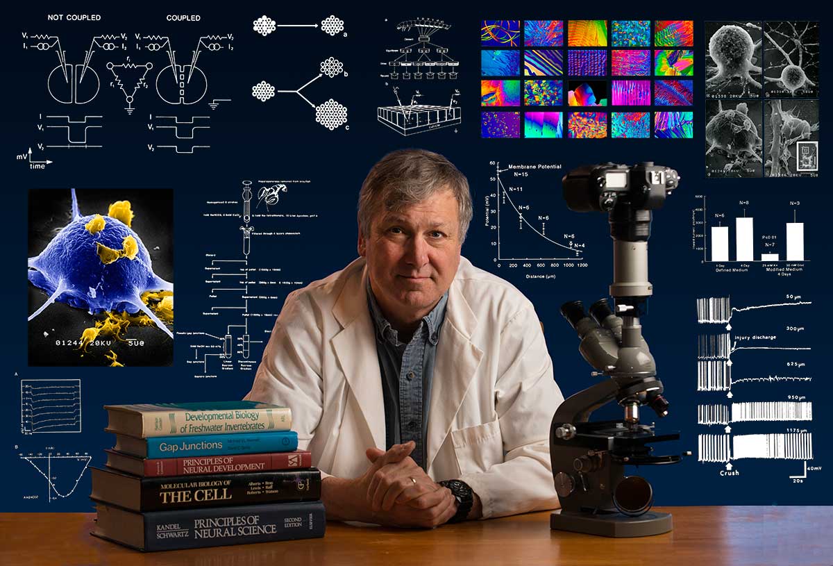
Biography: Robert Berdan is a professional nature photographer living in Calgary, AB specializing in nature, wildlife and science photography. Robert retired from Cell\Neurobiology research to pursue photography full time years ago. Robert offers photo instruction ain all aspects of nature photography, Photoshop training, photomicrography and macro-photography. Portrait of Robert by Dr. Sharif Galal showing some of Robert's research in the background.
Email at: rberdan@scienceandart.org
Web sites: www.canadiannaturephotographer.com www.scienceandart.org
Phone: MST 10 am -7 pm (403) 247-2457.